

Hematopoietic stem/progenitor involvement in retinal microvascular repair during diabetes: Implications for bone marrow rejuvenation
- PMID: 29042190
- PMCID: PMC6335030
- DOI: 10.1016/j.visres.2017.06.016
Hematopoietic stem/progenitor involvement in retinal microvascular repair during diabetes: Implications for bone marrow rejuvenation
Abstract
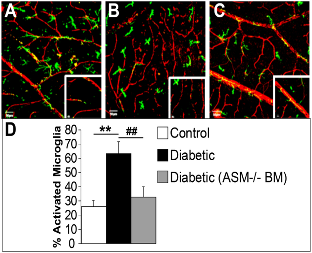
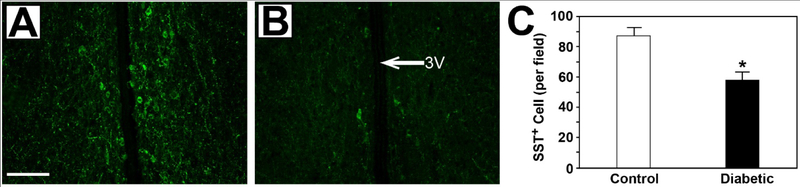
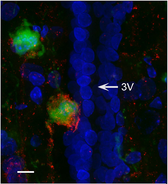
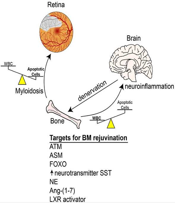
The widespread nature of diabetes affects all organ systems of an individual including the bone marrow. Long-term damage to the cellular and extracellular components of the bone marrow leads to a rapid decline in the bone marrow-hematopoietic stem/progenitor cells (HS/PCs) compartment. This review will highlight the importance of bone marrow microenvironment in maintaining bone marrow HS/PC populations and the contribution of these key populations in microvascular repair during the natural history of diabetes. The autonomic nervous system can initiate and propagate bone marrow dysfunction in diabetes. Systemic pharmacological strategies designed to protect the bone marrow-HS/PC population from diabetes induced-oxidative stress and advanced glycation end product accumulation represent a new approach to target diabetic retinopathy progression. Protecting HS/PCs ensures their participation in vascular repair and reduces the risk of vasogdegeneration occurring in the retina.
Keywords: Bone marrow microenvironment; Diabetic retinopathy; Hematopoietic stem cells.
Copyright © 2017. Published by Elsevier Ltd.
Conflict of interest statement
Disclosures
The authors declare that they have no conflicts of interest including any financial, personal or other relationships with other people or organizations within three years of beginning the submitted work that could inappropriately influence, or be perceived to influence, this work. All authors have materially participated in the research and/or article preparation of this article. All authors have approved the final article. All animal experiments comply with the
Figures
Similar articles
-
The Bone Marrow Microenvironment for Hematopoietic Stem Cells.Adv Exp Med Biol. 2017;1041:5-18. doi: 10.1007/978-3-319-69194-7_2. Adv Exp Med Biol. 2017. PMID: 29204826
-
Long-term diabetes impairs repopulation of hematopoietic progenitor cells and dysregulates the cytokine expression in the bone marrow microenvironment in mice.Basic Res Cardiol. 2010 Nov;105(6):703-12. doi: 10.1007/s00395-010-0109-0. Epub 2010 Jul 22. Basic Res Cardiol. 2010. PMID: 20652278
-
Diabetic retinopathy is associated with bone marrow neuropathy and a depressed peripheral clock.J Exp Med. 2009 Dec 21;206(13):2897-906. doi: 10.1084/jem.20090889. Epub 2009 Nov 23. J Exp Med. 2009. PMID: 19934019 Free PMC article.
-
Concise review: Current concepts in bone marrow microenvironmental regulation of hematopoietic stem and progenitor cells.Stem Cells. 2013 Jun;31(6):1044-50. doi: 10.1002/stem.1370. Stem Cells. 2013. PMID: 23509002 Free PMC article. Review.
-
Minireview: complexity of hematopoietic stem cell regulation in the bone marrow microenvironment.Mol Endocrinol. 2014 Oct;28(10):1592-601. doi: 10.1210/me.2014-1079. Epub 2014 Aug 1. Mol Endocrinol. 2014. PMID: 25083740 Free PMC article. Review.
Cited by
-
Preventing diabetic retinopathy by mitigating subretinal space oxidative stress in vivo.Vis Neurosci. 2020 Jun 15;37:E002. doi: 10.1017/S0952523820000024. Vis Neurosci. 2020. PMID: 32536351 Free PMC article. Review.
-
Bone Marrow CD133+ Stem Cells Ameliorate Visual Dysfunction in Streptozotocin-induced Diabetic Mice with Early Diabetic Retinopathy.Cell Transplant. 2018 Jun;27(6):916-936. doi: 10.1177/0963689718759463. Epub 2018 May 2. Cell Transplant. 2018. PMID: 29717657 Free PMC article.
-
Pericyte-Endothelial Interactions in the Retinal Microvasculature.Int J Mol Sci. 2020 Oct 8;21(19):7413. doi: 10.3390/ijms21197413. Int J Mol Sci. 2020. PMID: 33049983 Free PMC article. Review.
-
Pathophysiology of Diabetic Retinopathy: Contribution and Limitations of Laboratory Research.Ophthalmic Res. 2019;62(4):196-202. doi: 10.1159/000500026. Epub 2019 Jul 30. Ophthalmic Res. 2019. PMID: 31362288 Free PMC article. Review.
-
Transcriptomic Profile of Lin-Sca1+c-kit (LSK) cells in db/db mice with long-standing diabetes.BMC Genomics. 2024 Aug 12;25(1):782. doi: 10.1186/s12864-024-10679-3. BMC Genomics. 2024. PMID: 39134978 Free PMC article.
References
-
- Antonetti DA, Barber AJ, Bronson SK, Freeman WM, Gardner TW, Jefferson LS, Kester M, Kimball SR, Krady JK, LaNoue KF, Norbury CC, Quinn PG, Sandirasegarane L, Simpson IA, Group JDRC, Diabetic retinopathy: seeing beyond glucose-induced microvascular disease, Diabetes, 55 (2006) 2401–2411. - PubMed
-
- Klein BE, Overview of epidemiologic studies of diabetic retinopathy, Ophthalmic Epidemiol, 14 (2007) 179–183. - PubMed
-
- Cheung N, Mitchell P, Wong TY, Diabetic retinopathy, Lancet, 376 (2010) 124–136. - PubMed
-
- International Diabetes Federation. IDF Diabetes Atlas, 7 ed, 7 ed., International Diabetes Federation, Place Published, 2015.
Publication types
MeSH terms
Grants and funding
LinkOut - more resources
Full Text Sources
Other Literature Sources
Medical
