. 2018 Mar;138(3):697-703.
doi: 10.1016/j.jid.2017.09.031. Epub 2017 Oct 16.

The Physicochemical Basis of Clofazimine-Induced Skin Pigmentation

Affiliations

The Physicochemical Basis of Clofazimine-Induced Skin Pigmentation

Mikhail D Murashov et al. J Invest Dermatol. 2018 Mar.

Abstract

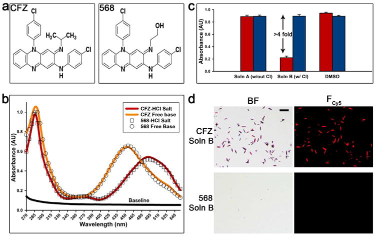
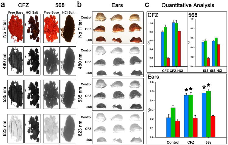
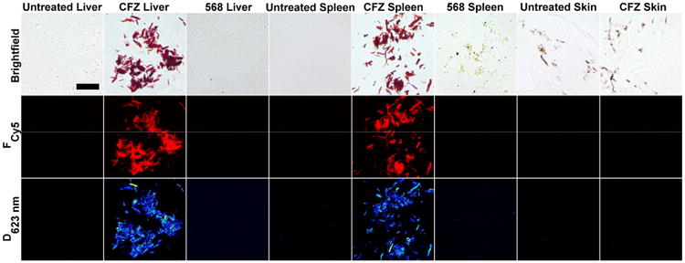
Clofazimine is a weakly basic, Food and Drug Administration-approved antibiotic recommended by the World Health Organization to treat leprosy and multi-drug-resistant tuberculosis. Upon prolonged treatment, clofazimine extensively bioaccumulates and precipitates throughout the organism, forming crystal-like drug inclusions (CLDIs). Due to the drug's red color, it is widely believed that clofazimine bioaccumulation results in skin pigmentation, its most common side effect. To test whether clofazimine-induced skin pigmentation is due to CLDI formation, we synthesized a closely related clofazimine analog that does not precipitate under physiological pH and chloride conditions that are required for CLDI formation. Despite the absence of detectable CLDIs in mice, administration of this analog still led to significant skin pigmentation. In clofazimine-treated mice, skin cryosections revealed no evidence of CLDIs when analyzed with a microscopic imaging system specifically designed for detecting clofazimine aggregates. Rather, the reflectance spectra of the skin revealed a signal corresponding to the soluble, free base form of the drug. Consistent with the low concentrations of clofazimine in the skin, these results suggest that clofazimine-induced skin pigmentation is not due to clofazimine precipitation and CLDI formation, but rather to the partitioning of the circulating, free base form of the drug into subcutaneous fat.


Conflict of interest statement

Conflict Of Interest: Gus R. Rosania is consulting for Bristol-Myers Squibb.

Figures

Figure 1
Figure 1. Comparison of CFZ and CFZ analog 568: structures, precipitation assay, and absorbance profiles
(a) The structural difference between CFZ and analog 568 at the tertiary amine is a specific substitution of the isopropyl group with a 2-hydroxyethyl. (b) The absorbance profiles (270-550nm) show the similarity of CFZ and analog 568 free base and hydrochloride salt forms, respectively. (c) The ability of both CFZ and analog 568 to precipitate as hydrochloride salt in simulated lysosomal conditions (pH 4.5). This was determined by measuring the absorbance (285nm) in different conditions: Soln A (no chloride), Soln B (100mM chloride), and DMSO (red = CFZ, blue = 568). (d) Hydrochloride salt precipitation in the presence of chloride (Soln B) was verified by brightfield (BF) and fluorescence microscopy (Fcy5). Scale bar = 50μm.
Figure 2
Figure 2. Optical properties of CFZ and CFZ analog 568 and quantitative analysis of skin pigmentation using various optical filters
(a) Images of CFZ and analog 568 free base and HCl salt powders and the extent of their reflectance using 480nm, 535nm, and 623nm optical filters. (b) Images of mouse ears after 3-4 weeks of CFZ or analog 568 treatment and the extent of their reflectance using 480nm, 535nm, and 623nm optical filters. (c) Quantitative analysis of the drug powder images (n=7) and images of mouse ears after 3-4 weeks of CFZ or 568 treatment compared to untreated mice (n=3) (blue = 480nm filter; green = 535nm filter; red = 623nm filter; *p < 0.01, ANOVA single factor, Tukey's HSD).
Figure 3
Figure 3. Multi-parameter imaging of CFZ and CFZ analog 568 treated and untreated tissues
Images of liver, spleen, and skin sections were taken using brightfield, Cy5 fluorescence (Fcy5), and dichroism at 623nm (D623nm) in order to identify and compare for CFZ-HCl/CLDI signatures with corresponding untreated samples. Scale Bar = 25μm.

References

    1. Arbiser JL, Moschella SL. Clofazimine: a review of its medical uses and mechanisms of action. J Am Acad Dermatol. 1995;32(2 Pt 1):241–7. - PubMed
    1. Arms AD, Travis CC Health USEPAOo, Assessment E. Reference Physiological Parameters in Pharmacokinetic Modeling. U.S. Environmental Protection Agency, Office of Health and Environmental Assessment; 1988.
    1. Baik J, Rosania GR. Molecular imaging of intracellular drug-membrane aggregate formation. Mol Pharm. 2011;8(5):1742–9. - PMC - PubMed
    1. Baik J, Rosania GR. Macrophages sequester clofazimine in an intracellular liquid crystal-like supramolecular organization. PLoS One. 2012;7(10):e47494. - PMC - PubMed
    1. Baik J, Stringer KA, Mane G, Rosania GR. Multiscale distribution and bioaccumulation analysis of clofazimine reveals a massive immune system-mediated xenobiotic sequestration response. Antimicrob Agents Chemother. 2013;57(3):1218–30. - PMC - PubMed

Publication types

LinkOut - more resources