

Calcium uptake and cytochrome c release from normal and ischemic brain mitochondria
- PMID: 29042253
- PMCID: PMC5964042
- DOI: 10.1016/j.neuint.2017.10.003
Calcium uptake and cytochrome c release from normal and ischemic brain mitochondria
Abstract
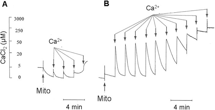
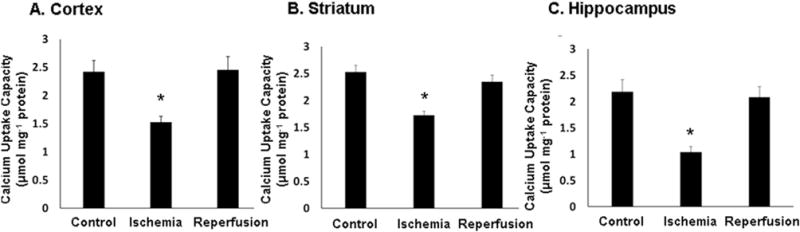
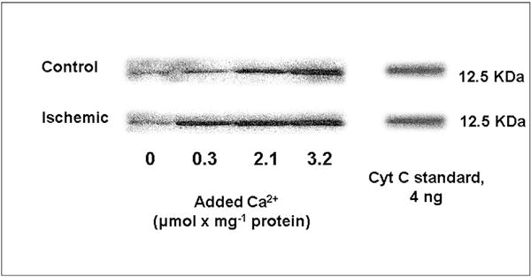
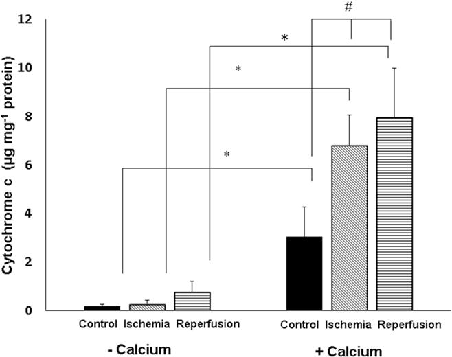
At abnormally elevated levels of intracellular Ca2+, mitochondrial Ca2+ uptake may compromise mitochondrial electron transport activities and trigger membrane permeability changes that allow for release of cytochrome c and other mitochondrial apoptotic proteins into the cytosol. In this study, a clinically relevant canine cardiac arrest model was used to assess the effects of global cerebral ischemia and reperfusion on mitochondrial Ca2+ uptake capacity, Ca2+ uptake-mediated inhibition of respiration, and Ca2+-induced cytochrome c release, as measured in vitro in a K+-based medium in the presence of Mg2+, ATP, and NADH-linked oxidizable substrates. Maximum Ca2+ uptake by frontal cortex mitochondria was significantly lower following 10 min cardiac arrest compared to non-ischemic controls. Mitochondria from ischemic brains were also more sensitive to the respiratory inhibition associated with accumulation of large levels of Ca2+. Cytochrome c was released from brain mitochondria in vitro in a Ca2+-dose-dependent manner and was more pronounced following both 10 min of ischemia alone and following 24 h reperfusion, in comparison to mitochondria from non-ischemic Shams. These effects of ischemia and reperfusion on brain mitochondria could compromise intracellular Ca2+ homeostasis, decrease aerobic and increase anaerobic cerebral energy metabolism, and potentiate the cytochrome c-dependent induction of apoptosis, when re-oxygenated mitochondria are exposed to abnormally high levels of intracellular Ca2+.
Keywords: Apoptosis; Cortex; Excitotoxicity; Hippocampus; Respiration.
Copyright © 2017 Elsevier Ltd. All rights reserved.
Figures
Similar articles
-
Therapeutic hypothermia attenuates global cerebral reperfusion-induced mitochondrial damage by suppressing dynamin-related protein 1 activation and mitochondria-mediated apoptosis in a cardiac arrest rat model.Neurosci Lett. 2017 Apr 24;647:45-52. doi: 10.1016/j.neulet.2017.02.065. Epub 2017 Feb 24. Neurosci Lett. 2017. PMID: 28242326
-
Ischemia-induced release of cytochrome c from mitochondria and up-regulation of Bcl-2 expression in rat hippocampus.Sheng Li Xue Bao. 2004 Apr 25;56(2):147-52. Sheng Li Xue Bao. 2004. PMID: 15127122
-
Mannosylated liposomal cytidine 5' diphosphocholine prevent age related global moderate cerebral ischemia reperfusion induced mitochondrial cytochrome c release in aged rat brain.Neuroscience. 2010 Dec 29;171(4):1287-99. doi: 10.1016/j.neuroscience.2010.09.049. Epub 2010 Sep 29. Neuroscience. 2010. PMID: 20883746
-
Targeting mitochondria for resuscitation from cardiac arrest.Crit Care Med. 2008 Nov;36(11 Suppl):S440-6. doi: 10.1097/ccm.0b013e31818a89f4. Crit Care Med. 2008. PMID: 20449908 Free PMC article. Review.
-
Astrocyte mitochondria in in vitro models of ischemia.J Bioenerg Biomembr. 2004 Aug;36(4):317-21. doi: 10.1023/B:JOBB.0000041761.61554.44. J Bioenerg Biomembr. 2004. PMID: 15377865 Review.
Cited by
-
The Effects of Potassium Cyanide on the Functional Recovery of Isolated Rat Hearts after Ischemia and Reperfusion: The Role of Oxidative Stress.Oxid Med Cell Longev. 2018 Jul 19;2018:5979721. doi: 10.1155/2018/5979721. eCollection 2018. Oxid Med Cell Longev. 2018. PMID: 30116485 Free PMC article.
-
Glial Cell AMPA Receptors in Nervous System Health, Injury and Disease.Int J Mol Sci. 2019 May 17;20(10):2450. doi: 10.3390/ijms20102450. Int J Mol Sci. 2019. PMID: 31108947 Free PMC article. Review.
-
Phytochemical, Antioxidant and Mitochondrial Permeability Transition Studies on Fruit-Skin Ethanol Extract of Annona muricata.J Toxicol. 2019 Dec 31;2019:7607031. doi: 10.1155/2019/7607031. eCollection 2019. J Toxicol. 2019. PMID: 32089679 Free PMC article.
-
MicroRNA-24 therapeutic potentials in infarction, stroke, and diabetic complications.Mol Biol Rep. 2024 Nov 9;51(1):1137. doi: 10.1007/s11033-024-10089-4. Mol Biol Rep. 2024. PMID: 39520600 Review.
-
Neuroprotective Effect of Taohong Siwu Decoction on Cerebral Ischemia/Reperfusion Injury via Mitophagy-NLRP3 Inflammasome Pathway.Front Pharmacol. 2022 Jun 8;13:910217. doi: 10.3389/fphar.2022.910217. eCollection 2022. Front Pharmacol. 2022. PMID: 35754465 Free PMC article.
References
-
- Andreyev A, Fahy B, Fiskum G. Cytochrome c release from brain mitochondria is independent of the mitochondrial permeability transition. FEBS Lett. 1998;439:373–376. - PubMed
-
- Andreyev A, Fiskum G. Calcium induced release of mitochondrial cytochrome c by different mechanisms selective for brain versus liver. Cell Death Differ. 1999;6:825–832. - PubMed
-
- Ankarcrona M, Dypbukt JM, Bonfoco E, Zhivotovsky B, Orrenius S, Lipton SA, Nicotera P. Glutamate-induced neuronal death: a succession of necrosis or apoptosis depending on mitochondrial function. Neuron. 1995;15:961–973. - PubMed
Publication types
MeSH terms
Substances
Grants and funding
LinkOut - more resources
Full Text Sources
Other Literature Sources
Miscellaneous
