Ruxolitinib-induced defects in DNA repair cause sensitivity to PARP inhibitors in myeloproliferative neoplasms
- PMID: 29042365
- PMCID: PMC5746670
- DOI: 10.1182/blood-2017-05-784942
Ruxolitinib-induced defects in DNA repair cause sensitivity to PARP inhibitors in myeloproliferative neoplasms
Abstract
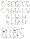
Myeloproliferative neoplasms (MPNs) often carry JAK2(V617F), MPL(W515L), or CALR(del52) mutations. Current treatment options for MPNs include cytoreduction by hydroxyurea and JAK1/2 inhibition by ruxolitinib, both of which are not curative. We show here that cell lines expressing JAK2(V617F), MPL(W515L), or CALR(del52) accumulated reactive oxygen species-induced DNA double-strand breaks (DSBs) and were modestly sensitive to poly-ADP-ribose polymerase (PARP) inhibitors olaparib and BMN673. At the same time, primary MPN cell samples from individual patients displayed a high degree of variability in sensitivity to these drugs. Ruxolitinib inhibited 2 major DSB repair mechanisms, BRCA-mediated homologous recombination and DNA-dependent protein kinase-mediated nonhomologous end-joining, and, when combined with olaparib, caused abundant accumulation of toxic DSBs resulting in enhanced elimination of MPN primary cells, including the disease-initiating cells from the majority of patients. Moreover, the combination of BMN673, ruxolitinib, and hydroxyurea was highly effective in vivo against JAK2(V617F)+ murine MPN-like disease and also against JAK2(V617F)+, CALR(del52)+, and MPL(W515L)+ primary MPN xenografts. In conclusion, we postulate that ruxolitinib-induced deficiencies in DSB repair pathways sensitized MPN cells to synthetic lethality triggered by PARP inhibitors.
© 2017 by The American Society of Hematology.
Conflict of interest statement
Conflict-of-interest disclosure: S.K. received honoraria and travel support for conferences from Novartis, Incyte, and Bristol-Myers Squibb. The remaining authors declare no competing financial interests.
Figures


Comment in
-
DNA distress creates lethal opportunity in MPN.Blood. 2017 Dec 28;130(26):2814-2816. doi: 10.1182/blood-2017-11-813006. Blood. 2017. PMID: 29284611 No abstract available.
Similar articles
-
Low JAK2 V617F Allele Burden in Ph-Negative Chronic Myeloproliferative Neoplasms Is Associated with Additional CALR or MPL Gene Mutations.Genes (Basel). 2021 Apr 12;12(4):559. doi: 10.3390/genes12040559. Genes (Basel). 2021. PMID: 33921387 Free PMC article.
-
MPL overexpression induces a high level of mutant-CALR/MPL complex: a novel mechanism of ruxolitinib resistance in myeloproliferative neoplasms with CALR mutations.Int J Hematol. 2021 Oct;114(4):424-440. doi: 10.1007/s12185-021-03180-0. Epub 2021 Jun 24. Int J Hematol. 2021. PMID: 34165774
-
Molecular genetics of BCR-ABL1 negative myeloproliferative neoplasms in India.Indian J Pathol Microbiol. 2018 Apr-Jun;61(2):209-213. doi: 10.4103/IJPM.IJPM_223_17. Indian J Pathol Microbiol. 2018. PMID: 29676359
-
Classical Philadelphia-negative myeloproliferative neoplasms: focus on mutations and JAK2 inhibitors.Med Oncol. 2018 Aug 3;35(9):119. doi: 10.1007/s12032-018-1187-3. Med Oncol. 2018. PMID: 30074114 Free PMC article. Review.
-
Changing concepts of diagnostic criteria of myeloproliferative disorders and the molecular etiology and classification of myeloproliferative neoplasms: from Dameshek 1950 to Vainchenker 2005 and beyond.Acta Haematol. 2015;133(1):36-51. doi: 10.1159/000358580. Epub 2014 Aug 7. Acta Haematol. 2015. PMID: 25116092 Review.
Cited by
-
JAK Inhibition for the Treatment of Myelofibrosis: Limitations and Future Perspectives.Hemasphere. 2020 Jul 21;4(4):e424. doi: 10.1097/HS9.0000000000000424. eCollection 2020 Aug. Hemasphere. 2020. PMID: 32903304 Free PMC article. Review.
-
Distinct effects of ruxolitinib and interferon-alpha on murine JAK2V617F myeloproliferative neoplasm hematopoietic stem cell populations.Leukemia. 2020 Apr;34(4):1075-1089. doi: 10.1038/s41375-019-0638-y. Epub 2019 Nov 15. Leukemia. 2020. PMID: 31732720 Free PMC article.
-
1H-Pyrazolo[3,4-b]quinolines: Synthesis and Properties over 100 Years of Research.Molecules. 2022 Apr 26;27(9):2775. doi: 10.3390/molecules27092775. Molecules. 2022. PMID: 35566124 Free PMC article. Review.
-
Management of myelofibrosis after ruxolitinib failure.Leuk Lymphoma. 2020 Aug;61(8):1797-1809. doi: 10.1080/10428194.2020.1749606. Epub 2020 Apr 16. Leuk Lymphoma. 2020. PMID: 32297800 Free PMC article.
-
PARP1 as a therapeutic target in acute myeloid leukemia and myelodysplastic syndrome.Blood Adv. 2021 Nov 23;5(22):4794-4805. doi: 10.1182/bloodadvances.2021004638. Blood Adv. 2021. PMID: 34529761 Free PMC article. Review.
References
-
- Lundberg P, Karow A, Nienhold R, et al. . Clonal evolution and clinical correlates of somatic mutations in myeloproliferative neoplasms. Blood. 2014;123(14):2220-2228. - PubMed
-
- Delic S, Rose D, Kern W, et al. . Application of an NGS-based 28-gene panel in myeloproliferative neoplasms reveals distinct mutation patterns in essential thrombocythaemia, primary myelofibrosis and polycythaemia vera. Br J Haematol. 2016;175(3):419-426. - PubMed
-
- Kubovcakova L, Lundberg P, Grisouard J, et al. . Differential effects of hydroxyurea and INC424 on mutant allele burden and myeloproliferative phenotype in a JAK2-V617F polycythemia vera mouse model. Blood. 2013;121(7):1188-1199. - PubMed
-
- Campbell PJ, Green AR. The myeloproliferative disorders. N Engl J Med. 2006;355(23):2452-2466. - PubMed
Publication types
MeSH terms
Substances
Grants and funding
LinkOut - more resources
Full Text Sources
Other Literature Sources
Molecular Biology Databases
Research Materials
Miscellaneous

