. 2017 Dec 8;292(49):20281-20291.
doi: 10.1074/jbc.M117.816751. Epub 2017 Oct 17.

Wilms tumor protein-dependent transcription of VEGF receptor 2 and hypoxia regulate expression of the testis-promoting gene Sox9 in murine embryonic gonads

Affiliations

Wilms tumor protein-dependent transcription of VEGF receptor 2 and hypoxia regulate expression of the testis-promoting gene Sox9 in murine embryonic gonads

Karin M Kirschner et al. J Biol Chem. .

Abstract

Wilms tumor protein 1 (WT1) has been implicated in the control of several genes in sexual development, but its function in gonad formation is still unclear. Here, we report that WT1 stimulates expression of Kdr, the gene encoding VEGF receptor 2, in murine embryonic gonads. We found that WT1 and KDR are co-expressed in Sertoli cells of the testes and somatic cells of embryonic ovaries. Vivo-morpholino-mediated WT1 knockdown decreased Kdr transcripts in cultured embryonic gonads at multiple developmental stages. Furthermore, WT1 bound to the Kdr promoter in the chromatin of embryonic testes and ovaries. Forced expression of the WT1(-KTS) isoform, which functions as a transcription factor, increased KDR mRNA levels, whereas the WT1(+KTS) isoform, which acts presumably on the post-transcriptional level, did not. ChIP indicated that WT1(-KTS), but not WT1(+KTS), binds to the KDR promoter. Treatment with the KDR tyrosine kinase inhibitor SU1498 or the KDR ligand VEGFA revealed that KDR signaling represses the testis-promoting gene Sox9 in embryonic XX gonads. WT1 knockdown abrogated the stimulatory effect of SU1498-mediated KDR inhibition on Sox9 expression. Exposure to 1% O2 to mimic the low-oxygen conditions in the embryo increased Vegfa expression but did not affect Sox9 mRNA levels in gonadal explants. However, incubation in 1% O2 in the presence of SU1498 significantly reduced Sox9 transcripts in cultured testes and increased Sox9 levels in ovaries. These findings demonstrate that both the local oxygen environment and WT1, which enhances KDR expression, contribute to sex-specific Sox9 expression in developing murine gonads.

Keywords: chromatin immunoprecipitation (ChiP); development; gene regulation; gene silencing; gene transcription; hypoxia; immunohistochemistry; ovary; testis; transcription factor.


Conflict of interest statement

The authors declare that they have no conflicts of interest with the contents of this article

Figures
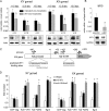
Figure 1.
Figure 1.
WT1 is necessary for normal Kdr gene expression in embryonic gonads and binds to the Kdr promoter. A, Kdr mRNA in cultured gonads of male (XY gonad) and female (XX gonad) mouse embryos with knockdown of WT1. The gonadal primordia were isolated from wild-type embryos at the indicated days dpc and incubated for 72 h in the presence of either mismatch or Wt1 antisense vivo-morpholino. The gonads of each embryo were used in a pairwise manner (i.e. the explant incubated with the mismatch vivo-morpholino served as a control for the Wt1 vivo-morpholino–treated gonad). Kdr and Actin transcripts were measured by RT-qPCR. Data representing means ± S.E. are shown as -fold differences relative to 11.5 dpc XY gonads treated with mismatch vivo-morpholino. *, p < 0.05, paired t test. Representative immunoblots below the graphs demonstrate knockdown of WT1 in the morpholino-treated gonads. B, Kdr mRNA levels in mesonephros-derived M15 cells transfected with either siRNA for WT1 knockdown (siWt1) or non-targeting siRNA (sicontrol). Kdr and Gapdh transcripts were quantified by real-time RT-qPCR. Values are shown as means ± S.E. (error bars), n = 4. Statistical significance is indicated by asterisks (paired t test; *, p < 0.05). WT1 and GAPDH proteins in M15 cells were detected by immunoblotting to determine knockdown efficiency, and representative data are shown. C and D, ChIP was performed to detect WT1 protein bound to the 5′-flanking region of the Kdr gene. The drawing (C) delineates the promoter, 5′-UTR, and translational start of the mouse Kdr gene and allocates the qPCR primers (F for forward and R for reverse) used for DNA amplification of the Kdr core promoter (F1/R1) and the 5′-UTR (F2/R2). The sequence of predicted WT1 binding sites is indicated. D, specific antibodies were chosen for immunoprecipitation of WT1 and histone (H3K4me3) proteins associated with chromatin in XY and XX embryonic gonads at 13.5 dpc. Normal rabbit IgG (nRbIgG) was used as a negative control. Note that immunoprecipitation with WT1 antibody enriches Kdr promoter DNA (F1/R1) in XY and XX gonads, whereas the 5′-UTR (F2/R2) is not enriched. Primers binding to a transcriptional inactive region of the genome were used as a negative control (ChIP neg). Enrichment of the Egr3 promoter served as a positive control. Data are presented as means ± S.E. (error bars) of -fold increase relative to the ChIP neg/nRbIgG control. Statistical significance is indicated by asterisks (*, p < 0.05, paired t test compared with ChIP neg control; n = 7).
Figure 2.
Figure 2.
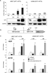
WT1(−KTS) stimulates KDR gene expression and binds to KDR chromatin. A, KDR mRNA levels in UB27 and UD28 cells expressing the WT1(−KTS) and WT1(+KTS) isoforms, respectively. The cells were grown in the presence of tetracycline (+tet) to keep WT1 expression suppressed. Removal of tetracycline from the cell culture medium (−tet) for the indicated periods increased WT1 proteins in UB27 and UD28 cells as detected by immunoblotting (bottom panels). KDR and GAPDH transcripts were measured by RT-qPCR. Values are shown as means ± S.E. (error bars); UB27 cells, n = 3; UD28 cells, n = 5. Statistical significance is indicated by asterisks (analysis of variance with Tukey's post-hoc test; F(3/11) = 5,959; *, p < 0.05). B and C, ChIP was performed to detect WT1 protein bound to the 5′-flanking region of the KDR gene. The drawing (B) delineates the promoter, 5′-UTR, and translational start of the human KDR gene and allocates the PCR primers (F for forward and R for reverse) used for DNA amplification. C, specific antibodies were chosen for immunoprecipitation of WT1 and histone proteins associated with chromatin in UB27 and UD28 cells. Note that KDR promoter DNA is enriched upon induction of WT1(−KTS) protein in UB27 cells (−tet), whereas induction of WT1(+KTS) protein in UD28 cells caused no enrichment. Analysis of chromatin isolated from non-induced UB27 and UD28 cells (+tet) as well as amplification of actin genomic DNA served as a negative control. Data (means ± S.E.) are presented as -fold increase relative to nRbIgG controls. Statistical significance is indicated by asterisks (*, p < 0.05, paired t test compared with nRbIgG; n = 4).
Figure 3.
Figure 3.
WT1 and KDR expression overlap in embryonic and adult gonads. Specific antibodies against WT1 (green) and KDR (red) were used to detect both proteins in the gonads of embryonic and adult rats. Immunolabeling identified Sertoli cells (>) and the coelomic epithelium (arrows in b and c) as the sites of WT1 expression in males. KDR is clearly co-expressed with WT1 in Sertoli cells (> in d and i). In the developing XX gonad, WT1 expression is observed in the somatic cells and later the pregranulosa cells. KDR is present at the basement membrane of the pregranulosa cells enclosing the late stage primary oocytes in the inner zone of the developing ovary (> in n). In adult ovaries, WT1 expression is observed in granulosa cells of the follicles (*). KDR is located at the outer boundary of the membrana granulosa (> in s). Incubation of the tissue sections with normal sera produced no specific staining signal (e, j, o, and t). Cell nuclei are stained with DAPI in some micrographs. Scale bars, 100 μm.
Figure 4.
Figure 4.
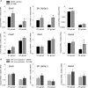
KDR signaling down-regulates key molecules for gonadal development in a WT1-dependent manner. A, gonadal explant cultures were established from XY and XX mouse embryos (12.5 dpc) and incubated with either KDR inhibitor SU1498 (10 μm) or vehicle (DMSO) for 48 h. Transcript levels of Sox9, Sf1 (Nr5a1), Amh, Fox2l, Dax1, and Gata4 were measured by RT-qPCR and normalized to Sdha transcripts. Data representing means ± S.E. (error bars) are shown as -fold differences compared with control (i.e. DMSO-treated XY gonads). Statistical significance is indicated by asterisks (paired t test; *, p < 0.05, n = 12 (XY) and n = 6 (XX)). B, gonadal explant cultures (XX and XY gonads, 12.5 dpc) were pretreated with Wt1 vivo-morpholino to knock down WT1 before incubation with the KDR inhibitor SU1498 (10 μm) or DMSO. Levels of Sox9, Sf1, and Gata4 mRNA were measured by RT-qPCR. Data representing means ± S.E. (error bars) are shown as -fold differences compared with control (i.e. DMSO-treated XY gonads), n = 9 (XY) and n = 6 (XX).
Figure 5.
Figure 5.
Repression of Sox9 by WT1 in XX gonads depends on KDR signaling. A, gonadal explant cultures (XX and XY gonads, 12.5 dpc) were treated with Wt1 vivo-morpholino, and the contralateral gonad of each embryo was incubated with mismatch vivo-morpholino as a control. Sox9 mRNA levels were measured by RT-PCR and normalized to Sdha transcripts. Data representing means ± S.E. (error bars) are shown as -fold differences compared with control (mismatch vivo-morpholino) XY gonads. Statistical significance is indicated by asterisks (paired t test; **, p < 0.01; ***, p < 0.005; n = 13 (XY) and n = 12 (XX)). B, gonadal explant cultures (XX and XY gonads, 12.5 dpc) were incubated with the KDR inhibitor SU1498 (10 μm) before treatment with Wt1 vivo-morpholino or mismatch vivo-morpholino. Sox9 mRNA levels were measured by RT-qPCR. Data representing means ± S.E. (error bars) are shown as -fold differences compared with control (mismatch vivo-morpholino) XY gonads. Statistical significance is indicated by asterisks (paired t test; *, p < 0.05; n = 7 (XY) and n = 8 (XX)).
Figure 6.
Figure 6.
Hypoxia, WT1, and VEGF/KDR signaling jointly regulate Sox9 expression in cultured murine embryonic gonads. A, the gonads of each embryo were treated separately for 48 h with either recombinant mouse VEGFA (rmVEGFA, 500 ng/ml) or bovine serum albumin as a negative control (ctrl.), n = 13 (XY) and n = 9 (XX). Sox9 mRNA levels were measured by RT-qPCR. Data representing means ± S.E. (error bars) are shown as -fold differences compared with control (mismatch vivo-morpholino) XY gonads. Statistical significance is indicated by asterisks (paired t test; *, p < 0.05). B–D, the gonads of each embryo (12.5 dpc) were incubated ex vivo for 24 h in 21% O2 (normoxia; Nx) followed by another 24 h in either 1% O2 (hypoxia; Hx) or (the second gonad) 21% O2 (Nx). Vegfa, Wt1, and Sox9 mRNA levels were measured by RT-qPCR and normalized to Sdha transcripts. Data representing means ± S.E. (error bars) are shown as -fold differences compared with levels in XY gonads in normoxia. Statistical significance is indicated by asterisks (paired t test; *, p < 0.05; ***, p < 0.005; n = 8 (XY) and n = 8 (XX)). E and F, gonadal explant cultures (XX and XY gonads, 12.5 dpc) were treated with the KDR inhibitor SU1498 (10 μm) (E) or SU1498 + Wt1 vivo-morpholino (F) before cultivation in hypoxia (1% O2) or normoxia (21% O2). Sox9 mRNA levels were measured by RT-qPCR and normalized to Sdha transcripts. Data representing means ± S.E. (error bars) are shown as -fold differences compared with levels in XY gonads in normoxia. Statistical significance is indicated by asterisks (paired t test; **, p < 0.01; ***, p < 0.005; n = 8 (XY) and n = 6 (XX)).

Similar articles

Cited by

References

    1. Koopman P., Gubbay J., Vivian N., Goodfellow P., and Lovell-Badge R. (1991) Male development of chromosomally female mice transgenic for Sry. Nature 351, 117–121 - PubMed
    1. Hacker A., Capel B., Goodfellow P., and Lovell-Badge R. (1995) Expression of Sry, the mouse sex determining gene. Development 121, 1603–1614 - PubMed
    1. Albrecht K. H., and Eicher E. M. (2001) Evidence that Sry is expressed in pre-Sertoli cells, and Sertoli and granulosa cells have a common precursor. Dev. Biol. 240, 92–107 - PubMed
    1. Bullejos M., and Koopman P. (2001) Spatially dynamic expression of Sry in mouse genital ridges. Dev. Dyn. 221, 201–205 - PubMed
    1. Chassot A. A., Gregoire E. P., Magliano M., Lavery R., and Chaboissier M. C. (2008) Genetics of ovarian differentiation: Rspo1, a major player. Sex. Dev. 2, 219–227 - PubMed

MeSH terms

LinkOut - more resources