Comparative Study
. 2017 Dec 15;292(50):20558-20569.
doi: 10.1074/jbc.M117.811398. Epub 2017 Oct 17.

Plant pathogenesis-related proteins of the cacao fungal pathogen Moniliophthora perniciosa differ in their lipid-binding specificities

Affiliations
Comparative Study

Plant pathogenesis-related proteins of the cacao fungal pathogen Moniliophthora perniciosa differ in their lipid-binding specificities

Rabih Darwiche et al. J Biol Chem. .

Abstract

Moniliophthora perniciosa is the causative agent of witches' broom disease, which devastates cacao cultures in South America. This pathogenic fungus infects meristematic tissues and derives nutrients from the plant apoplast during an unusually long-lasting biotrophic stage. To survive, the fungus produces proteins to suppress the plant immune response. Proteins of the PR-1 (pathogenesis-related 1)/CAP superfamily have been implicated in fungal virulence and immune suppression. The genome of M. perniciosa encodes 11 homologues of plant PR-1 proteins, designated MpPR-1 proteins, but their precise mode of action is poorly understood. In this study, we expressed MpPR-1 proteins in a yeast model lacking endogenous CAP proteins. We show that some members of the MpPR-1 family bind and promote secretion of sterols, whereas others bind and promote secretion of fatty acids. Lipid binding by purified MpPR-1 occurs with micromolar affinity and is saturable in vitro Sterol binding by MpPR-1 requires the presence of a flexible loop region containing aromatic amino acids, the caveolin-binding motif. Remarkably, MpPR-1 family members that do not bind sterols can be converted to sterol binders by a single point mutation in the caveolin-binding motif. We discuss the possible implications of the lipid-binding activity of MpPR-1 family members with regard to the mode of action of these proteins during M. perniciosa infections.

Keywords: fatty acid; fatty acid binding protein; infection; lipid-protein interaction; sterol.


Conflict of interest statement

The authors declare that they have no conflicts of interest with the contents of this article

Figures

Figure 1.
Figure 1.
Some MpPR-1 family members complement the sterol export defect of yeast mutants lacking Pry function. A, heme and Say1 deficient double mutant cells (hem1Δ say1Δ) containing either no plasmid (−), an empty plasmid (Empty), or a plasmid-borne copy of Pry1 or of the indicated MpPR-1 family member were cultivated in the presence of radiolabeled [14C]cholesterol. Lipids were extracted from the cell pellet (P) and the culture supernatant (S), separated by TLC, and visualized by phosphorimaging. The position of free cholesterol (FC), CA, steryl esters (STE) and unidentified lipids (*) are indicated to the right. The experiment was repeated five times with similar results. B, quantification of the export of CA. The export index indicates the relative levels of CA exported by the cells (the ratio between extracellular CA and the sum of intracellular and extracellular CA). The data represent the means ± S.D. of five independent experiments. Asterisks denote statistical significance of the export phenotype relative to hem1Δ say1Δ double mutant cells. ***, p < 0.0001. n.s., not significant.
Figure 2.
Figure 2.
Comparison of the CBM of the 11 MpPR-1 family members from M. perniciosa. The positions of aromatic amino acids (ø) within the CBM are indicated at the top. Residues in red squares at position 3 of the motif mark the amino acids that were mutated. The aromatic amino acid in position 8 of the CBM is conserved only in few family members. Proteins selected for further analysis are highlighted in yellow: MpPR-1d, MpPR-1e, MpPR-1i, and MpPR-1k.
Figure 3.
Figure 3.
Single point mutations in the CBM convert MpPR-1s that do not export sterols into sterol exporters. A, substitutions of aromatic amino acids in the third position of the CBM convert the sterol exporting MpPR-1d and MpPR-1k into non-exporting mutant versions (MpPR-1d→e and MpPR-1k→i). Conversely, introduction of an aromatic residue at position 3 of the CBM results in the conversion of the non-sterol exporting MpPR-1e and MpPR-1i into gain of function versions (MpPR-1e→d and MpPR-1i→k). hem1Δ say1Δ double mutant cells expressing the indicated wild-type (wt) or mutant version of MpPR-1 proteins were radiolabeled with [14C]cholesterol. Lipids were extracted from the cell pellet (P) and the culture supernatant (S) and separated by TLC. The position of free cholesterol (FC), CA, steryl esters (STE), and an unidentified lipid (*) are indicated to the right. The experiment was repeated three times with similar results. B, the export index was plotted as the ration between exported to total CA. The data represent the means ± S.D. of three independent experiments. Asterisks denote statistical significance of the export phenotype. ***, p < 0.0001.
Figure 4.
Figure 4.
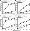
MpPR-1 bind cholesterol in vitro. A–D, MpPR-1 family members that export sterols in vivo also bind sterols in vitro. Sterol binding by the indicated MpPR-1 wild-type (wt) protein and the respective mutant versions was assessed using 100 pmol of purified protein and an increasing concentration of [3H]cholesterol (0–400 pmol). The protein was separated from unbound ligand by adsorption to an anion-exchange matrix, and bound radioligand was quantified by scintillation counting. Background binding was recorded by performing the binding assay in the absence of protein. The data represent the means ± S.D. of three independent experiments. n.d., not determined.
Figure 5.
Figure 5.
MpPR-1d binds cholesteryl acetate, sitosterol, and ergosterol in vitro. Binding specificity of MpPR-1d was assessed in a competition binding assay in which an unlabeled sterol competes with [3H]cholesterol for binding to the protein. Purified MpPR-1d protein (100 pmol) was incubated with 50 pmol of [3H]cholesterol and either an equal concentration (50 pmol) or an excess (500 pmol) of the indicated sterol. The data represent the means ± S.D. of two independent experiments. Asterisks denote statistical significance relative to the control containing only the radiolabeled cholesterol and purified MpPR-1d. **, p < 0.001; *, p < 0.01. chol, cholesterol.
Figure 6.
Figure 6.
MpPR-1 family members that do not export sterols export fatty acids. A, wild-type (wt), faa1Δ faa4Δ double mutant and pry1Δ pry2Δ faa1Δ faa4Δ quadruple mutant cells expressing the indicated MpPR-1 family members were cultivated in minimal medium overnight at 30 °C. Lipids were then extracted from the cell pellet, and the culture supernatant and fatty acids were quantified by GC-MS. The values are plotted as export index representing the amount of exported fatty acids relative to total fatty acids present in the cell pellet and the supernatant. The data represent the means ± S.D. of three independent experiments. Asterisks denote statistical significance relative to the faa1Δ faa4Δ double mutant (***, p < 0.0001; **, p < 0.001; *, p < 0.01). n.s., not significant. B, MpPR-1 family members that do not export fatty acids do not provide the essential function of CAP proteins in cells that export fatty acids. Wild-type and quintuple mutant cells lacking Pry function and the acyl-CoA synthetases Faa1 and Faa4 expressing a plasmid borne copy of either Pry1 or the indicated MpPR-1 family member were serially diluted 10-fold and spotted on plates with the indicated medium. The URA3-marked plasmid-borne copy of FAA1 present in these cells cannot be lost on 5-fluoroorotic acid (5-FOA) medium in cells expressing MpPR-1d or MPPR-1k, the two MpPR-1 family members that do not export or bind fatty acids.
Figure 7.
Figure 7.
MpPR-1 family members that export fatty acids in vivo also bind fatty acids in vitro. A, fatty acid binding to the indicated MpPR-1 family members was assessed by an in vitro binding assay with purified protein (100 pmol) and an increasing concentration of [3H]palmitic acid (0–400 pmol). The protein was separated from unbound ligand by adsorption to an anion-exchange matrix, and the bound radioligand was quantified by scintillation counting. Background binding was recorded by performing the binding assay in the absence of protein. B, CAP family members can bind both sterols and fatty acids simultaneously. Radioligand binding by yeast Pry1 and the respective gain of function versions of MpPR-1, MpPR-1e→d, and MpPR-1i→k was assessed using 100 pmol of purified protein and either [3H]cholesterol (50 pmol), [3H]palmitic acid (50 pmol), or both [3H]cholesterol and [3H]palmitic acid (each at 50 pmol) in the binding reaction. The data represent the means ± S.D. of three independent experiments.
Figure 8.
Figure 8.
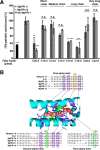
MpPR-1 family members bind saturated long chain and polyunsaturated fatty acids. A, fatty acid-binding specificity was assessed by a competition binding assay in which an equal concentration (50 pmol) of the unlabeled fatty acid of the indicated chain length and degree of unsaturation competes with the radiolabeled [3H]palmitic acid (50 pmol) for binding to the protein (100 pmol). The data represent the means ± S.D. of three independent experiments. Asterisks denote statistical significance relative to the control containing only the radiolabeled palmitic acid and purified MpPR-1i or MpPR-1e, respectively. ***, p < 0.0001; **, p < 0.001. n.s., not significant. B, sequence alignment around the fatty acid-binding pocket. The sequence of tablysin-15, yeast Pry1, and the M. perniciosa MpPR-1 family members analyzed in more detail are shown. The numbering on the top corresponds to the amino acid positions in Pry1. Valine at position 254 of Pry1 (boxed in green) is important for fatty acid binding (19). Key residues shown in colors are those forming the fatty acid binding pocket in tablysin-15 (17). B was adapted from Ref. .

Similar articles

Cited by

References

    1. Meinhardt L. W., Rincones J., Bailey B. A., Aime M. C., Griffith G. W., Zhang D., and Pereira G. A. (2008) Moniliophthora perniciosa, the causal agent of witches' broom disease of cacao: what's new from this old foe. Mol. Plant Pathol. 9, 577–588 - PMC - PubMed
    1. Teixeira P. J., Thomazella D. P., and Pereira G. A. (2015) Time for chocolate: current understanding and new perspectives on cacao witches' broom disease research. PLoS Pathog. 11, e1005130. - PMC - PubMed
    1. Aime M. C., and Phillips-Mora W. (2005) The causal agents of witches' broom and frosty pod rot of cacao (chocolate, Theobroma cacao) form a new lineage of Marasmiaceae. Mycologia 97, 1012–1022 - PubMed
    1. Teixeira P. J., Thomazella D. P., Reis O., do Prado P. F., do Rio M. C., Fiorin G. L., José J., Costa G. G., Negri V. A., Mondego J. M., Mieczkowski P., and Pereira G. A. (2014) High-resolution transcript profiling of the atypical biotrophic interaction between Theobroma cacao and the fungal pathogen Moniliophthora perniciosa. Plant Cell 26, 4245–4269 - PMC - PubMed
    1. Barau J., Grandis A., Carvalho V. M., Teixeira G. S., Zaparoli G. H., do Rio M. C., Rincones J., Buckeridge M. S., and Pereira G. A. (2015) Apoplastic and intracellular plant sugars regulate developmental transitions in witches' broom disease of cacao. J. Exp. Bot. 66, 1325–1337 - PMC - PubMed

Publication types

MeSH terms