Review
. 2018 Feb 7;13(2):300-317.
doi: 10.2215/CJN.00620117. Epub 2017 Oct 17.

Thrombotic Microangiopathy and the Kidney

Affiliations
Review

Thrombotic Microangiopathy and the Kidney

Vicky Brocklebank et al. Clin J Am Soc Nephrol. .

Abstract

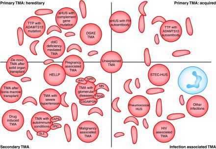
Thrombotic microangiopathy can manifest in a diverse range of diseases and is characterized by thrombocytopenia, microangiopathic hemolytic anemia, and organ injury, including AKI. It can be associated with significant morbidity and mortality, but a systematic approach to investigation and prompt initiation of supportive management and, in some cases, effective specific treatment can result in good outcomes. This review considers the classification, pathology, epidemiology, characteristics, and pathogenesis of the thrombotic microangiopathies, and outlines a pragmatic approach to diagnosis and management.

Keywords: Acute Kidney Injury; Anemia; Complement; Hemolytic; Purpura; Thrombotic Microangiopathies; Thrombotic Thrombocytopenic; atypical hemolytic uremic syndrome; glomerular disease; kidney.


Figures

Figure 1.
Figure 1.
Thrombotic microangiopathies are classified into: Inherited or acquired primary; secondary; or infection associated TMAs. Current classifications define primary TMAs as hereditary (mutations in ADAMTS13, MMACHC (cb1c deficiency), or genes encoding complement proteins) or acquired (autoantibodies to ADAMTS13, or autoantibodies to complement FH, which is associated with homozygous CFHR3/1 deletion). TMA is associated with various infections: in STEC-HUS and pneumococcal HUS, distinct mechanisms result in TMA; in other infections, the processes are not defined and in some cases the infection may trigger manifestation of a primary TMA. Secondary TMAs occur in a spectrum of conditions, and in many cases the pathogenic mechanisms are multifactorial or unknown. The classification presented here is not unequivocal: in some secondary TMAs, for example pregnancy-associated TMA or de novo TMA after transplantation, a significant proportion of individuals will have a genetic predisposition to a primary TMA. AAV, ANCA-associated vasculitis; ADAMTS13, a disintegrin and metalloproteinase with a thrombospondin type 1 motif, member 13; aHUS, atypical hemolytic uremic syndrome; C3G, C3 glomerulopathy; CAPS, catastrophic antiphospholipid syndrome; cblC, cobalamin C type; DGKE, gene encoding diacylglycerol kinase ε; FH, factor H; HELLP, syndrome of hemolysis, elevated liver enzymes, and low platelets; HUS, hemolytic uremic syndrome; IgAN, IgA nephropathy; MN, membranous nephropathy; MPGN, membranoproliferative GN; SRC, scleroderma renal crisis; STEC, shiga toxin–producing Escherichia coli; TMA, thrombotic microangiopathy; TTP, thrombotic thrombocytopenic purpura.
Figure 2.
Figure 2.
The pathological features of thrombotic microangiopathies reflect the tissue responses to endothelial injury. (A) Glomerular paralysis with capillary loops containing abundant erythrocytes (silver, ×400). (B) Thrombus in an artery (hematoxylin and eosin, ×400). (C) Glomerular capillary lumina containing fibrin thrombi (red) and erythrocytes (yellow) (Martius Scarlet Blue, ×400). (D) Erythrocyte fragments within the arterial vessel wall (arrow) (hematoxylin and eosin, ×400). (E) Mucoid thickening and obliteration of the lumen of a small artery (hematoxylin and eosin, ×400). (F) Myxoid intimal thickening of small artery (hematoxylin and eosin, ×400). (G) Fibrinoid necrosis of arterial wall (Martius Scarlet Blue, ×400). (H) Reduplication of glomerular basement membrane (arrow) and fibrillary mesangium (periodic acid–Schiff, ×400). (I) Glomerulus with endothelial swelling and erythrocyte fragmentation (arrow) (hematoxylin and eosin, ×400). (J) Electron micrograph demonstrating fibrin tactoids (black) in glomerular capillary (×10,000).
Figure 3.
Figure 3.
A diagnostic algorithm for the investigation and management of a patient presenting with thrombotic microangiopathy. Supportive treatment is essential. This comprises fluid management and, if indicated, judicious packed red blood cell transfusion, intensive care unit admission, and RRT. Platelet transfusion may worsen the TMA and so should be avoided if possible. The complete evaluation of a patient presenting with TMA comprises complement analysis and investigations for all conditions in which TMA can manifest, and usually enables the disease cause to be established. However, collating the results of the requisite investigations may take considerable time. The clinical evaluation during the acute presentation of a TMA requires some time-critical decision-making, which should focus initially on the consideration of TTP, because immediate management is imperative given the high mortality rate if untreated, and therefore in adults, PE should be instituted on the presumption that it is TTP unless other evidence is available that strongly suggests an alternative cause. In children, in whom TTP is rarer, first-line treatment with eculizumab should be considered if complement-mediated aHUS is suspected and should not be delayed while ADAMTS13 activity is determined. In the absence of a defined cause, complement-mediated aHUS is presumed and treatment with eculizumab is recommended, pending the complete evaluation. Eculizumab is not universally available; in these circumstances, treatment should comprise PE, and there may be a role for liver transplantation. *Full complement evaluation is recommended (Kidney Disease: Improving Global Outcomes consensus [6]) in individuals with pregnancy-associated aHUS and de novo transplantation-associated aHUS because of the high prevalence of rare genetic variants described in these subgroups, and in cases of STEC-HUS that result in ESRD, as rare genetic variants have been described after HUS recurrence in a subsequent kidney transplant. Additionally, it is recognized that infection can trigger manifestation of complement-mediated aHUS. In other secondary cases of aHUS, insufficient evidence exists to recommend a full genetic evaluation, although rare genetic variants have been described in many of these presentations. In cases where the role of complement is as yet unclear, the clinician should decide on the evaluation on the basis of the potential clinical consequences of a positive result (e.g., listing for renal transplantation). +ve, positive; AAV, ANCA-associated vasculitis; Ab, antibody; ACA, anticentromere antibody; ACEI, angiotensin-converting enzyme inhibitor; ADAMTS13, a disintegrin and metalloproteinase with a thrombospondin type 1 motif, member 13; Ag, antigen; aHUS, atypical hemolytic uremic syndrome; ANA antinuclear antibody; anti-ds DNA, antidouble-stranded DNA; anti–Scl70, anti-topoisomerase I antibody; BMT; bone marrow transplant; C3G, C3 glomerulopathy; CAPS, catastrophic antiphospholipid syndrome; DGKE, gene encoding diacylglycerol kinase ε; DIC, disseminated intravascular coagulation; FH, factor H; FI, factor I; Hb, hemoglobin; HUS, hemolytic uremic syndrome; LDH, lactate dehydrogenase; MAHA, microangiopathic hemolytic anemia; MN, membranous nephropathy; MPGN, membranoproliferative GN; PE, plasma exchange; SRC, scleroderma renal crisis; STEC, shiga toxin–producing Escherichia coli; Stx, shiga toxin; T-Ag, Thomsen–Friedenreich antigen; TMA, thrombotic microangiopathy; TTP, thrombotic thrombocytopenic purpura.
Figure 4.
Figure 4.
Unfettered complement activation ultimately results in thrombus formation, platelet consumption, vascular occlusion and mechanical hemolysis. Complement is activated by the alternative, classic, and lectin pathways. The alternative pathway of complement is a positive amplification loop. C3b interacts with factor B, which is then cleaved by factor D to form the C3 convertase C3bBb. Unchecked, this leads to activation of the terminal complement pathway with generation of the effector molecules, the anaphylatoxin C5a and the membrane attack complex (C5b-9). To protect host cells from bystander damage, the alternative pathway is downregulated by complement regulators including factor H (FH), factor I (FI), and CD46. In complement-mediated aHUS, activating mutations in C3 and CFB and loss-of-function mutations in CFH, CFI, and CD46, in addition to autoantibodies to FH, result in overactivation of the alternative pathway. This leads to immune cell and platelet activation and endothelial cell damage and swelling, with consequent thrombus formation, platelet consumption, vascular occlusion, and mechanical hemolysis. There is evidence that complement is activated in other TMAs, but whether this is a causative, disease modifier, or bystander phenomenon has not yet been elucidated. Eculizumab is a humanized mAb that binds to C5 and prevents activation of the terminal pathway; it thereby inhibits the generation of the effector molecules that cause TMA in individuals in whom a primary defect in complement underlies the TMA pathogenesis. Its role in the treatment of other TMAs is undefined. aHUS, atypical hemolytic uremic syndrome; TMA, thrombotic microangiopathy.

References

    1. Moake JL: Thrombotic microangiopathies. N Engl J Med 347: 589–600, 2002 - PubMed
    1. George JN, Nester CM: Syndromes of thrombotic microangiopathy. N Engl J Med 371: 654–666, 2014 - PubMed
    1. Kerr H, Richards A: Complement-mediated injury and protection of endothelium: Lessons from atypical haemolytic uraemic syndrome. Immunobiology 217: 195–203, 2012 - PMC - PubMed
    1. Nester CM, Barbour T, de Cordoba SR, Dragon-Durey MA, Fremeaux-Bacchi V, Goodship TH, Kavanagh D, Noris M, Pickering M, Sanchez-Corral P, Skerka C, Zipfel P, Smith RJ: Atypical aHUS: State of the art. Mol Immunol 67: 31–42, 2015 - PubMed
    1. Scully M, Cataland S, Coppo P, de la Rubia J, Friedman KD, Kremer Hovinga J, Lammle B, Matsumoto M, Pavenski K, Sadler E, Sarode R, Wu H; International Working Group for Thrombotic Thrombocytopenic Purpura: Consensus on the standardization of terminology in thrombotic thrombocytopenic purpura and related thrombotic microangiopathies. J Thromb Haemost 15: 312–322, 2017. - PubMed

Publication types

MeSH terms

Substances