. 2017 Oct 17;8(1):1000.
doi: 10.1038/s41467-017-01068-4.

Cis P-tau is induced in clinical and preclinical brain injury and contributes to post-injury sequelae

Affiliations

Cis P-tau is induced in clinical and preclinical brain injury and contributes to post-injury sequelae

Onder Albayram et al. Nat Commun. .

Abstract

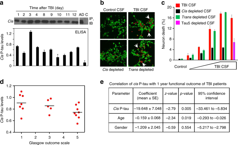
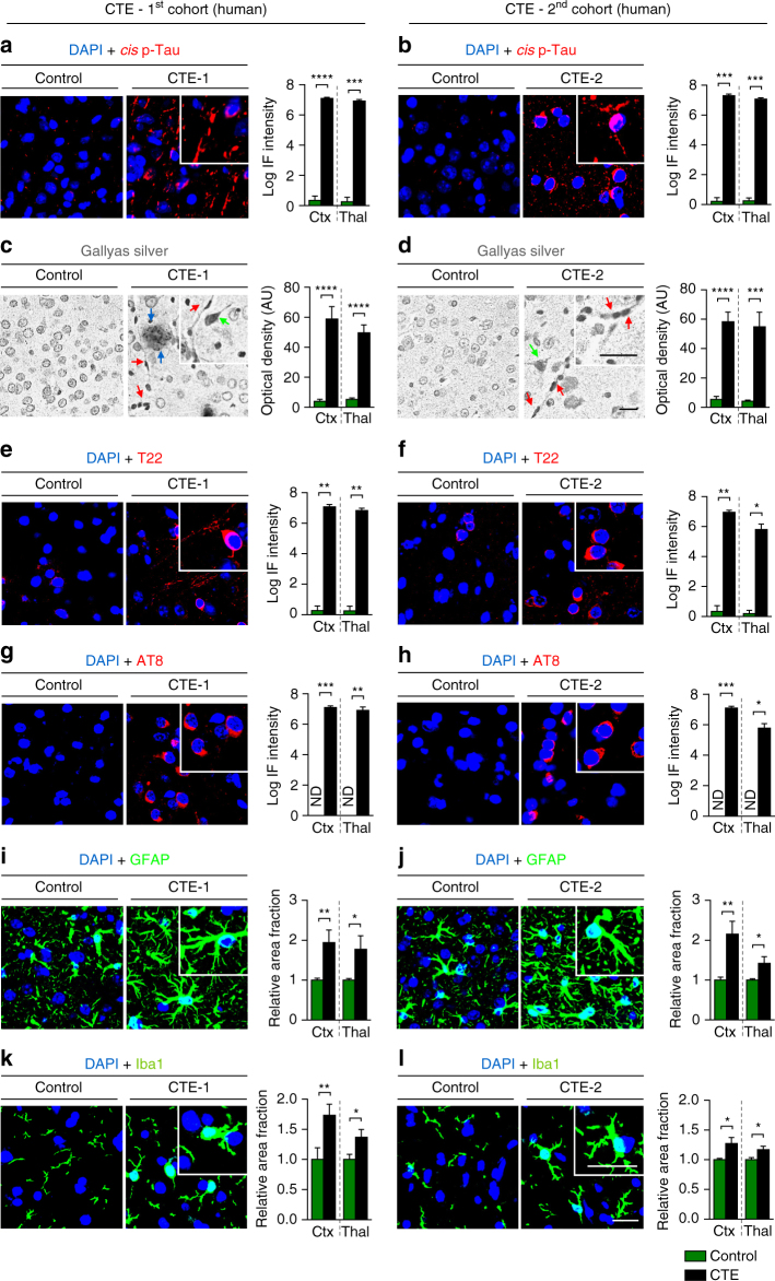
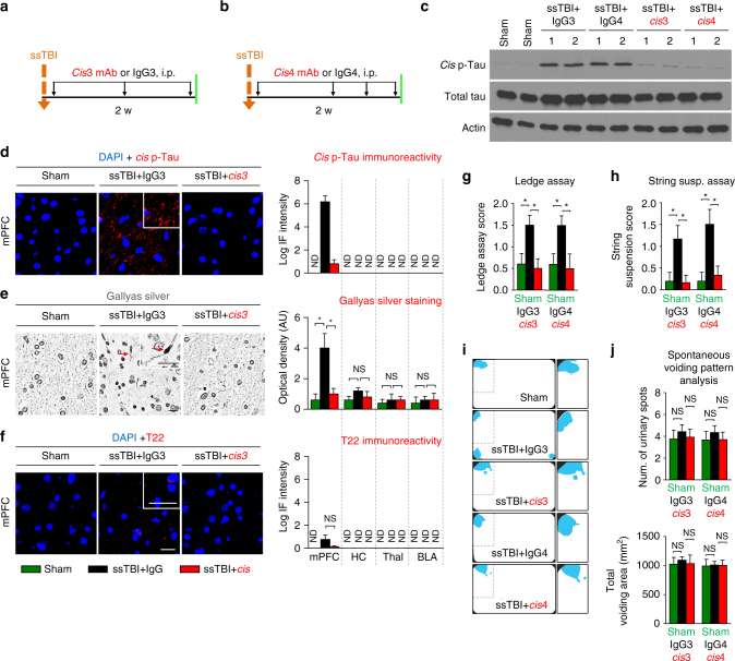
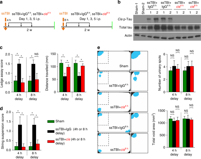
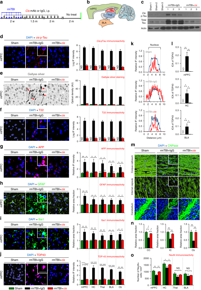
Traumatic brain injury (TBI) is characterized by acute neurological dysfunction and associated with the development of chronic traumatic encephalopathy (CTE) and Alzheimer's disease. We previously showed that cis phosphorylated tau (cis P-tau), but not the trans form, contributes to tau pathology and functional impairment in an animal model of severe TBI. Here we found that in human samples obtained post TBI due to a variety of causes, cis P-tau is induced in cortical axons and cerebrospinal fluid and positively correlates with axonal injury and clinical outcome. Using mouse models of severe or repetitive TBI, we showed that cis P-tau elimination with a specific neutralizing antibody administered immediately or at delayed time points after injury, attenuates the development of neuropathology and brain dysfunction during acute and chronic phases including CTE-like pathology and dysfunction after repetitive TBI. Thus, cis P-tau contributes to short-term and long-term sequelae after TBI, but is effectively neutralized by cis antibody treatment.Induction of the cis form of phosphorylated tau (cis P-tau) has previously been shown to occur in animal models of traumatic brain injury (TBI), and blocking this form of tau using antibody was beneficial in a rodent model of severe TBI. Here the authors show that cis P-tau induction is a feature of several different forms of TBI in humans, and that administration of cis P-tau targeting antibody to rodents reduces or delays pathological features of TBI.


Conflict of interest statement

Dr Lu and Dr Zhou are inventors of Pin1 technology including patents and patent application on tau antibodies, which was licensed by BIDMC to Pinteon Therapeutics. Both Dr Lu and Dr Zhou own equity in, and consult for, Pinteon. Their interests were reviewed and are managed by BIDMC in accordance with its conflict of interest policy. The remaining authors declare no competing financial interests.

Figures

Fig. 1
Fig. 1
Severe TBI in humans due to motor vehicle accidents, assaults or falls induces prominent axonal injury and axonal cis P-tau induction in the cortex. a, b Severe TBI in humans induces early cis P-tau induction and axonal injury in the cortex. Whereas neither cis nor trans P-tau nor axonal injury was detected in normal brains or 1 h after TBI due to motor vehicle accident, robust cis P-tau and axonal injury, but not trans P-tau were detected in the cortex, but not in the hippocampus, as early as 8 h after motor vehicle accident, as detected by double IF, followed by isotype-specific secondary antibodies a or Gallyas silver staining b. Microscope images correspond to the cortex and hippocampus of control and TBI patients. TBI cases due to falls and assaults are shown in Supplementary Fig. 1. The number of TBI patients is 14. White arrows point to cis P-tau localization to axons; Red arrows point to axonal bulb. White scale bars, 20 µm and black scale bars, 40 µm. c, d cis P-tau (red) is diffusely co-localized (white arrows) with the axon marker tau (green) c, but not the dendrite marker MAP2 (green) d in human TBI cortex, with little cis in control, as detected by double IF, followed by confocal microscopy. eh Cis P-tau is robustly induced after TBI in the absence of tau oligomers or tangles. Cortical sections of severe human TBI due to motor vehicle accidents were doubly immunostained with cis mAb (red) and T22 (tau oligomers) e, g or AT100 (tau tangles) f, h, followed by confocal microscopy. Normal controls as well as CTE and AD brains were used as negative and positive controls, respectively. Of note, cis mAb partially co-localized with T22 or AT100 in CTE or AD brains, but not in acute TBI brains
Fig. 2
Fig. 2
Cis P-tau in the CSF of severe TBI patients is neurotoxic and correlates with clinical outcome at 1 year after injury. a Detection of cis P-tau in the CSF of a TBI patient was performed by subjecting control CSF or CSF specimens collected from EVD at different times after TBI due to jumping into an oncoming train to immunoprecipitation with cis mAb, followed by immunoblotting with rabbit anti-tau mAb (top panel) or to direct ELISA with cis mAb in OD at 450 nm (bottom panel). Post-mortem Alzheimer’s patient CSF (AD) and control CSF were used as positive and negative controls, respectively. b, c Addition of TBI CSF to neurons induces dose-dependent cell death in recipient cells, which are fully rescued by immunodepletion with cis, but not trans mAb. Human control and TBI CSF specimens were added to culture media of SY5Y neurons for 3 days, followed by the live (green)/dead (red, white arrows) cell assay. For immunodepletion, cis or trans mAb was incubated with TBI CSF to deplete cis or trans p-tau before adding to cells. n = 3. d, e Cis P-tau levels of CSFs taken day 4 to 6 days after injury of acute TBI patients were measured by direct ELISA and an ordered logistic regression was used to model 1 year GOS as the outcome and cis P-tau level in OD at 450 nm as the main predictor, controlling for age, gender and initial Glasgow Coma Scale. Supplementary Table 2 shows demographic information for the subjects. n = 20
Fig. 3
Fig. 3
Cis P-tau is expressed in both the cortex and deep brain regions and correlates with various neuropathological features in CTE patients. CTE brain sections of the cortex and thalamus from eight collision sport athletes under ages of 75 from two independent sources and age-matched controls (Supplementary Table 3) were subjected to immunostaining or Gallyas silver staining, followed by confocal microscopy and light microscopy, respectively, to detect cis P-tau and various CTE pathologies. Cis P-tau staining a, b was correlated with the presence of axonal pathology, as detected by Gallyas silver staining c, d, tau oligomerization (T22) e, f, early tangles (AT8) g, h, astrogliosis (GFAP) i, j and microgliosis (Iba1) k, l in two different brain regions. Inset images are the high magnification image of selected area denoted by the white. Of note, there was not detectable signals when secondary antibodies were used alone (data not shown). Scale bar, 40 μm. Different types of pathologies in CTE brains revealed by Gallyas silver staining: senile plaque (blue arrow); neurofibrillary tangles (green arrow); axonal bulb (red arrow). Ctx, parasagittal cortex; Thal, thalamus; ND, not detectable; NS, not significant. Images were quantified and results are shown as means ± S.E.M. and p-values calculated using unpaired two-tailed parametric Student’s t-test. *p < 0.05, **p < 0.01, ***p < 0.001, ****p < 0.0001
Fig. 4
Fig. 4
Eliminating cis P-tau in ssTBI mice with cis mAb prevents a range of pathological and functional outcomes during the acute phases. Mice were subjected to one of two treatment regimens involving 3 or 4 i.p. injections of cis mAb for 2-weeks a, b, followed by functional, biochemical, and pathological examinations. Orange arrows, ssTBI; black arrows, antibody injection; green lines, functional, or pathological assays. Cis mAb treatment of ssTBI mice for 10 days effectively eliminated cis P-tau induction and total tau accumulation, as detected by immunostaining and immunoblotting c, d, restored axonal pathology by Gallyas silver staining e, without any tau oligomerization f, as well as prevented sensorimotor coordination deficits, as detected by Ledge assay g and string suspension h at 2 weeks after injury, with no change in urinary pattern at this time (that is, all mice urinated in the corner of cages indicated by blue fluorescent urine, as expected for normal mice) i, j. Microscope images correspond to the medial prefrontal cortex of sham (left), ssTBI + IgG (middle), and ssTBI + cis mAb (right) with quantification data in different brain regions being present at right panels. Inset images are the high magnification image of selected area denoted by the white. Scale bar, 40 μm. Axonal bulb also referred to as a retraction ball indicated with red arrow in Gallyas silver staining. mPFC, medial prefrontal cortex; HC, hippocampus; Thal, thalamus; BLA, basolateral amygdala. ND, not detectable; NS, not significant. Brains from 4−5 WT male mice were studied in immunohistochemistry, 5−6 mice underwent urinary pattern test and 9−10 WT mice underwent other behavioral studies per group. The data were presented as means ± SEM. The p-values were calculated using unpaired two-tailed parametric Student’s t-test. *p < 0.05, **p < 0.01, ***p < 0.001, ****p < 0.0001
Fig. 5
Fig. 5
Eliminating cis P-tau in ssTBI mice with cis mAb prevents a range of pathological and functional outcomes during the chronic phase. Mice were subjected to long-term treatment regimens over 4-months, followed by a 2-month washout a and then underwent functional, biochemical, and pathological examinations. Orange arrows, ssTBI; black arrows, antibody injection; green lines, functional, or pathological assays. Cis mAb treatment of ssTBI mice for 4 months effectively eliminated cis P-tau induction and total tau accumulation b, c, and prevented the development of axonal pathology d and tau oligomerization e both in the cortex and hippocampus, as well as prevented sensorimotor coordination deficits, as detected by Ledge assay f and string suspension g and urinary incontinence as assayed by spontaneous urinary pattern i at 6 months after injury, with no deficit in novel object location recognition h. Microscope images correspond to the medial prefrontal cortex of sham (left), ssTBI + IgG (middle), and ssTBI + cis mAb (right) with quantification data in different brain regions being present at right panels. Inset images are the high magnification image of selected area denoted by the white. Scale bar, 40 μm. Axonal bulb also referred to as a retraction ball indicated with red arrow in Gallyas silver staining. mPFC, medial prefrontal cortex; HC, hippocampus; Thal, thalamus; BLA, basolateral amygdala. ND, not detectable; NS, not significant. Brains from 4−5 WT male mice were studied in immunohistochemistry, 5−6 mice underwent urinary pattern test and 9−10 WT mice underwent other behavioral studies per group. The data were presented as means ± SEM. The p-values were calculated using unpaired two-tailed parametric Student’s t-test. *p < 0.05, **p < 0.01, ***p < 0.001, ****p < 0.0001
Fig. 6
Fig. 6
Treating ssTBI mice with 3 i.p. cis mAb with 4 or 8 h delay eliminates cis P-tau and restores sensorimotor coordination deficits at 2 weeks after injury. Treating ssTBI mice with 3 i.p. of cis mAb even with 4 or 8 h delay a effectively eliminated cis P-tau induction b, and restored sensorimotor coordination deficits (ledge assay and string suspension assay) c, d. Urinary incontinence was not observed after 2-weeks e. Orange arrows, ssTBI; black arrows, antibody injection; green arrows, functional, or pathological assays. The data were presented as means ± SEM. The p-values were calculated using unpaired two-tailed parametric Student’s t-test. *p < 0.05, NS, not significant
Fig. 7
Fig. 7
Eliminating cis P-tau in rmTBI mice with cis mAb prevents the development of a range of pathological features resembling those found in human CTE. Mice were subjected to seven mild TBI events over 9 days and were treated with cis mAb or IgG isotype control over 4 months, followed by 2 months of washout, before assaying pathologies in different brain regions a, b. Blue arrows, rmTBI; black arrows, antibody injection; green line, functional, or pathological assays. Cis mAb treatment of rmTBI mice eliminated induction and spreading of cis P-tau and total tau c, d, prevented the development and spreading of axonal pathology e, tau oligomerization f, APP accumulation g, GFAP-positive astrocyte h, Iba-positive microglia i, TDP-43 pathology with increased cytoplasmic mislocalization of TDP-43 jl. Line graphs showing the relative IF intensity of TDP-43 across a single cell k, l (Blue line, DAPI; red line, TDP-43). Cis mAb treatment also prevented the demyelination as detected by CNPase IF m, n, and neuronal loss o in different brain regions. Shorter exposure of ECL for total tau immunoblots in c was used due to a huge increase in total tau in rmTBI mice, as expected because cis P-tau is resistant to protein degradation. Microscope images corresponded to the medial prefrontal cortex of sham (left), rmTBI + IgG (middle), and rmTBI + cis mAb (right) with quantification data in different brain regions being present at right panels. Inset images are the high magnification image of selected area denoted by the white. Scale bar, 40 μm. Red arrows point to axonal bulb in Gallyas silver staining. mPFC, medial prefrontal cortex; HC, hippocampus; Thal, thalamus; BLA, basolateral amygdala; CC, corpus callosum; IC, internal capsule; Cb, cerebellum. ND, not detectable; NS, not significant. Brains from 4−5 WT male mice were studied in immunohistochemistry per group. The data are presented as means ± SEM. The p-values were calculated using unpaired two-tailed parametric Student’s t-test. *p < 0.05, **p < 0.01, ***p < 0.001, ****p < 0.0001
Fig. 8
Fig. 8
Eliminating cis P-tau in rmTBI mice with cis mAb prevents the development of clinically relevant functional deficits. Cis mAb treatment of rmTBI mice prevents sensorimotor coordination deficits, as detected by Ledge assay a, string suspension b and accelerated rotarod c, and urinary incontinence, as assayed by spontaneous urinary pattern analysis d, e and memory deficit, as assayed by novel object location recognition test at 6 months after injury f, g. 5−6 mice underwent urinary pattern test and 9−10 WT mice underwent other behavioral studies per group. The data are presented as means ± SEM. The p-values were calculated using unpaired two-tailed parametric Student’s t-test. *p < 0.05, **p < 0.01, ***p < 0.001, ****p < 0.0001
Fig. 9
Fig. 9
A model for the roles of cis P-tau and its mAb in the development and treatment of ssTBI and rmTBI. ssTBI or rmTBI causes persistent and robust cis P-tau induction before other tau pathology likely due to axon injury. Cis P-tau mainly localizes to axons and causes and spreads axonal pathology, contributing to the development and progression of a range of neuropathological and functional outcomes during acute and chronic phases, including those pathological features resembling human CTE. Treatment of ssTBI or rmTBI mice with cis mAb not only eliminates cis P-tau and blocks its spreading, but also prevents the development and progression of a range of neuropathological and functional outcomes after injury

Similar articles

Cited by

References

    1. Faul, M., Xu, L., Wald, M. M. & Coronado, V. G. Traumatic brain injury in the United States: emergency department visits, hospitalizations, and deaths, 2002–2006. Centers for Disease Control and Prevention [online], http://www.cdc.gov/traumaticbraininjury/tbi_ed.html (2010).
    1. Langlois JA, Rutland-Brown W, Wald MM. The epidemiology and impact of traumatic brain injury: a brief overview. J. Head. Trauma. Rehabil. 2006;21:375–378. doi: 10.1097/00001199-200609000-00001. - DOI - PubMed
    1. Center for Disease Control Nonfatal traumatic brain injuries from sports and recreation activities --- United States, 2001--2005. MMWR. 2007;56:733–737. - PubMed
    1. Weinberger S. Bombs’ hidden impact: the brain war. Nature. 2011;477:390–393. doi: 10.1038/477390a. - DOI - PubMed
    1. Miller G. The invisible wounds of war. Healing the brain, healing the mind. Science. 2011;333:514–517. doi: 10.1126/science.333.6042.514. - DOI - PubMed

Publication types

MeSH terms