

β-defensin 1 expression in HCV infected liver/liver cancer: an important role in protecting HCV progression and liver cancer development
- PMID: 29042578
- PMCID: PMC5645372
- DOI: 10.1038/s41598-017-13332-0
β-defensin 1 expression in HCV infected liver/liver cancer: an important role in protecting HCV progression and liver cancer development
Abstract
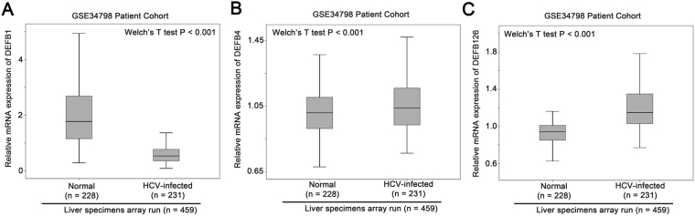
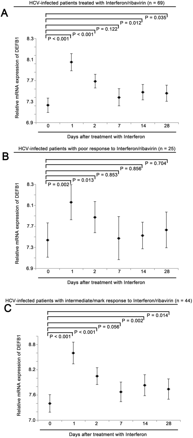
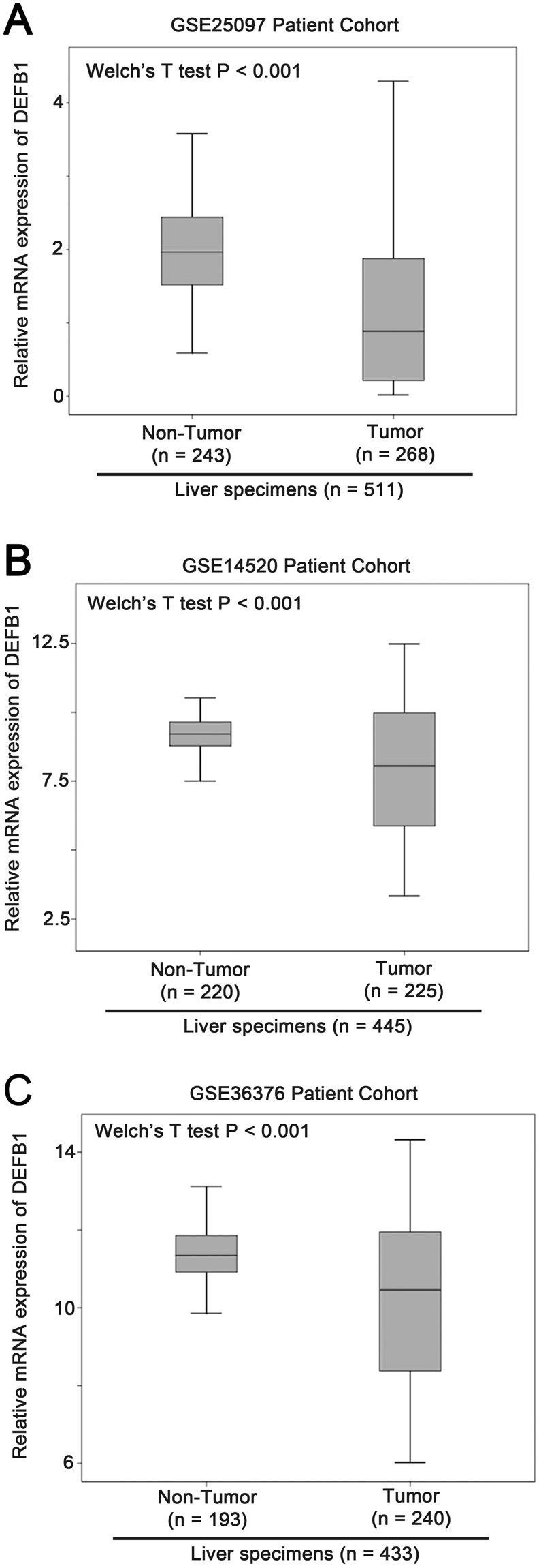
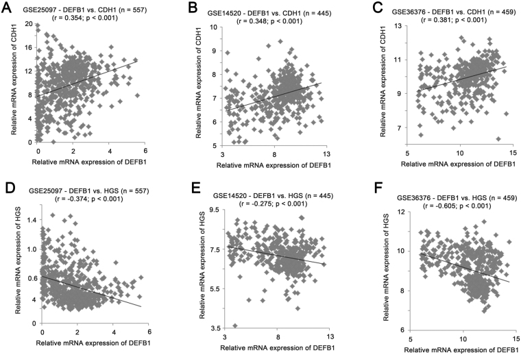
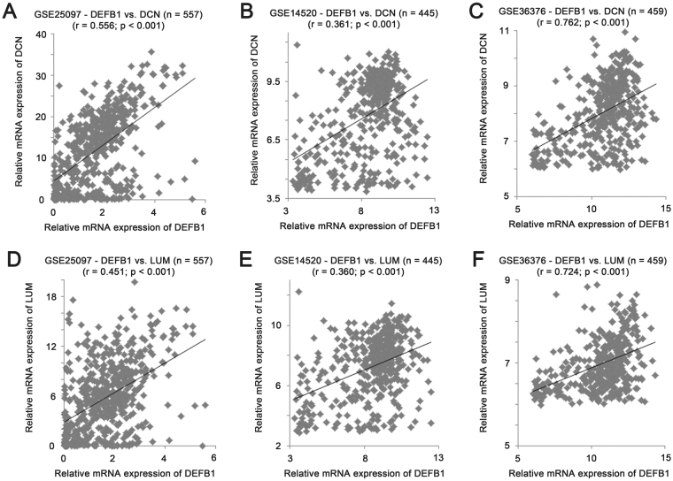
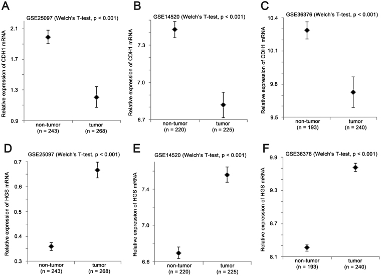
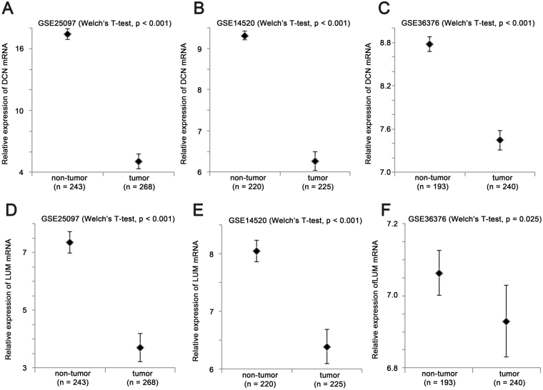
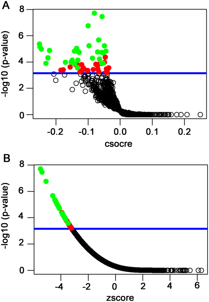
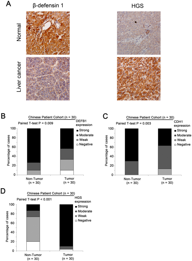
β-defensin family plays a role in host defense against viral infection, however its role in HCV infection is still unknown. In this study, we demonstrated that β-defensin 1 was significantly reduced in HCV-infected liver specimens. Treatment with interferon and ribavirin upregulated β-defensin-1, but not other β-defensin tested, with the extent and duration of upregulation associated with treatment response. We investigated β-defensin family expression in liver cancer in publicly available datasets and found that among all the β-defensins tested, only β-defensin 1 was significantly downregulated, suggesting β-defensin 1 plays a crucial role in liver cancer development. Further analysis identified E-cadherin as the top positive correlated gene, while hepatocyte growth factor-regulated tyrosine kinase substrate as the top negative correlated gene. Expression of two proteoglycans were also positively correlated with that of β-defensin 1. We have also identified small molecules as potential therapeutic agents to reverse β-defensin 1-associated gene signature. Furthermore, the downregulation of β-defensin 1 and E-cadherin, and upregulation of hepatocyte growth factor-regulated tyrosine kinase substrate, were further confirmed in liver cancer and adjacent normal tissue collected from in-house Chinese liver cancer patients. Together, our results suggest β-defensin 1 plays an important role in protecting HCV progression and liver cancer development.
Conflict of interest statement
The authors declare that they have no competing interests.
Figures
Similar articles
-
Increased miR-16 expression induced by hepatitis C virus infection promotes liver fibrosis through downregulation of hepatocyte growth factor and Smad7.Arch Virol. 2015 Aug;160(8):2043-50. doi: 10.1007/s00705-015-2474-3. Epub 2015 Jun 13. Arch Virol. 2015. PMID: 26071245
-
MicroRNA let-7g cooperates with interferon/ribavirin to repress hepatitis C virus replication.J Mol Med (Berl). 2016 Mar;94(3):311-20. doi: 10.1007/s00109-015-1348-1. Epub 2015 Oct 21. J Mol Med (Berl). 2016. PMID: 26489607
-
Insights into the pathobiology of hepatitis C virus-associated cirrhosis: analysis of intrahepatic differential gene expression.Am J Pathol. 2002 Feb;160(2):641-54. doi: 10.1016/S0002-9440(10)64884-5. Am J Pathol. 2002. PMID: 11839585 Free PMC article.
-
HCV-hepatocellular carcinoma: new findings and hope for effective treatment.Microsc Res Tech. 2005 Nov;68(3-4):130-48. doi: 10.1002/jemt.20227. Microsc Res Tech. 2005. PMID: 16276514 Review.
-
Hepatitis C: From inflammatory pathogenesis to anti-inflammatory/hepatoprotective therapy.World J Gastroenterol. 2018 Dec 21;24(47):5297-5311. doi: 10.3748/wjg.v24.i47.5297. World J Gastroenterol. 2018. PMID: 30598575 Free PMC article. Review.
Cited by
-
Etiology of viral induced acute liver failure and defensins as potential therapeutic agents in ALF treatment.Front Immunol. 2023 Apr 21;14:1153528. doi: 10.3389/fimmu.2023.1153528. eCollection 2023. Front Immunol. 2023. PMID: 37153560 Free PMC article. Review.
-
Defensins: A Double-Edged Sword in Host Immunity.Front Immunol. 2020 May 7;11:764. doi: 10.3389/fimmu.2020.00764. eCollection 2020. Front Immunol. 2020. PMID: 32457744 Free PMC article. Review.
-
Expression and Function of Host Defense Peptides at Inflammation Sites.Int J Mol Sci. 2019 Dec 22;21(1):104. doi: 10.3390/ijms21010104. Int J Mol Sci. 2019. PMID: 31877866 Free PMC article. Review.
-
Expression of the human antimicrobial peptide β-defensin-1 is repressed by the EGFR-ERK-MYC axis in colonic epithelial cells.Sci Rep. 2018 Dec 21;8(1):18043. doi: 10.1038/s41598-018-36387-z. Sci Rep. 2018. PMID: 30575780 Free PMC article.
-
Defensins: Exploring Their Opposing Roles in Colorectal Cancer Progression.Cancers (Basel). 2024 Jul 23;16(15):2622. doi: 10.3390/cancers16152622. Cancers (Basel). 2024. PMID: 39123348 Free PMC article. Review.
References
Publication types
MeSH terms
Substances
LinkOut - more resources
Full Text Sources
Other Literature Sources
Medical
