. 2017 Oct 17;7(1):13409.
doi: 10.1038/s41598-017-13728-y.

Developmental stage-dependent regulation of spine formation by calcium-calmodulin-dependent protein kinase IIα and Rap1

Affiliations

Developmental stage-dependent regulation of spine formation by calcium-calmodulin-dependent protein kinase IIα and Rap1

Solveigh Cornelia Koeberle et al. Sci Rep. .

Abstract

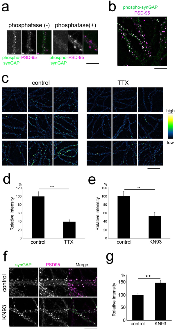
The roles of calcium-calmodulin-dependent protein kinase II-alpha (CaMKIIα) in the expression of long-term synaptic plasticity in the adult brain have been extensively studied. However, how increased CaMKIIα activity controls the maturation of neuronal circuits remains incompletely understood. Herein, we show that pyramidal neurons without CaMKIIα activity upregulate the rate of spine addition, resulting in elevated spine density. Genetic elimination of CaMKIIα activity specifically eliminated the observed maturation-dependent suppression of spine formation. Enhanced spine formation was associated with the stabilization of actin in the spine and could be reversed by increasing the activity of the small GTPase Rap1. CaMKIIα activity was critical in the phosphorylation of synaptic Ras GTPase-activating protein (synGAP), the dispersion of synGAP from postsynaptic sites, and the activation of postsynaptic Rap1. CaMKIIα is already known to be essential in learning and memory, but our findings suggest that CaMKIIα plays an important activity-dependent role in restricting spine density during postnatal development.


Conflict of interest statement

The authors declare that they have no competing interests.

Figures

Figure 1
Figure 1
Spine densities of wild-type and CaMKIIα KI hippocampal neurons maintained in slice cultures. (a) Representative images of oblique, apical, and basal dendrites at different developmental stages. Scale bars, 5 µm. (bd) Quantitative analysis of spine density in hippocampal pyramidal neurons maintained in slice cultures of wild-type and CaMKIIα KI mice at 9/10 DIV, 16/17 DIV, 23/24 DIV and 30/31 DIV. Spine density was measured from images of oblique (b), apical (c), and basal (d) branches of dendrites, collected from 7–22 experiments. Data are presented as the mean ± SEM, n = 7–22 cells, *p < 0.05, **p < 0.01 or ***p < 0.001, two-way ANOVA followed by Tukey’s post-hoc test. (e) The protein expression of CaMKIIα at 10, 13, 16 and 22 DIV in slices from KI and wild-type mice. CaMKIIα protein expression was low at 10 DIV in both KI and wild-type mice. A large increase in protein expression was observed from 10 to 16 DIV. Full-size images of the gels and blots are shown in Supplementary Fig. 1.
Figure 2
Figure 2
Spine formation and elimination in CA1 pyramidal neurons in CaMKIIα KI and wild-type hippocampal slices. (a and b) Spines formed (arrows at Day2) and eliminated (arrows at Day1) in the oblique branches of CA1 pyramidal neurons in mature slices (19–23 DIV), as detected by time-lapse imaging of GFP-expressing cells over 24 h. The dendritic segments of wild-type (a) and KI (b) slices are presented. Scale bars, 5 µm. (c) Quantitative analysis of spine formation and elimination in wild-type and KI mice. Data are presented as the mean ± SEM, n = 5 cells from 2 independent cultures for wild-type mice, n = 5 cells from 3 independent cultures for KI mice, *p < 0.05, t-test. (d) Inhibiting AMPA receptors with CNQX (20 µM), and NMDA receptors with AP-V (50 µM) for 4 days (from 19 to 23 DIV) reduced the spine density of KI neurons to wild-type neuron levels. Control spine densities were based on the same set of the data shown in Fig. 1. Data are presented as the mean ± SEM, n = 21 and 37 cells from 10 and 11 independent cultures of WT and KI control, n = 10 and 9 cells from 3 independent cultures of WT and KI APV treat ment, n = 13 and 23 cells from 4 independent cultures of WT and KI CNQX treat ment, ***p < 0.001, t-test.
Figure 3
Figure 3
Regulation of spine dynamics by CaMKII in dissociated neurons. (a) Time-lapse imaging of spines in dissociated hippocampal neurons expressing RFP and treated with the CaMKII inhibitor KN93 or its inactive analogue KN92. Gain and loss of spines are marked by arrows. Bar, 5 µm. (b) Enhancement of the spine turnover rate in neurons treated with KN93. Data are presented as the mean ± SEM, (neurons at 19–20 DIV, n = 12 cells from 3 independent cultures), ***p < 0.001, **p < 0.01, t-test. (c) Time-lapse imaging of spines in dissociated hippocampal neurons expressing RFP along with GFP-tagged wild-type CaMKII or the kinase-dead mutant of CaMKII (K42R). Newly formed spines are marked by arrows. Bar, 5 µm. (d) Pseudocolour images of neurons expressing GFP-tagged wild-type CaMKIIα or the K42R mutant at similar levels. Bar, 5 µm. (e) Quantification of the expression level of GFP-tagged wild-type CaMKIIα or the K42R mutant. Data are presented as the mean ± SEM (neurons at 14–16 DIV, n = 11 cells from 3 independent cultures for wild-type CaMKIIa, n = 20 cells from 3 independent cultures for K42R CaMKIIα), n.s. p > 0.05, t-test. (f) Quantification of the increase in spine density from 14 to 16 DIV in neurons expressing GFP-tagged wild-type CaMKIIα or the K42R mutant. Data are presented as the mean ± SEM, (neurons at 14–16 DIV, n = 11 cells from 3 independent cultures for wild-type CaMKIIa, n = 20 cells from 3 independent cultures for K42R CaMKIIα), **p < 0.01, t-test.
Figure 4
Figure 4
CaMKII-dependent regulation of Rap1 activity. (a) Quantification of active Rap1 and active Ras immunoreactivity in the dendrites of CaMKIIα KI or wild-type neurons. Data are presented as the mean ± SEM, (neurons at 21–22 DIV, Rap1; n = 42 and 27 cells from 4 independent cultures of wild-type and CaMKIIα KI hippocampi, Ras; n = 32 and 33 cells from 5 independent cultures of wild-type and CaMKIIα KI hippocampi), *p < 0.05, t-test. (b) Immunoreactivity of active Rap1 in the dendrites of hippocampal neurons at 22–23 DIV and immunostaining for the presynaptic marker vGluT1 or the postsynaptic marker synGAP. Active Rap1-positive punctae are either colocalized with (arrows) or separated from (arrowheads) the synaptic markers. Bar, 5 µm. (c) Immunocytochemistry of KN93-treated neurons exposed to the anti-active Rap1 antibody. The intensity of active-Rap1 punctae at synGAP-positive postsynaptic sites was lower in neurons treated with KN93 (arrows). Bar, 5 µm. (d) Quantification of active Rap1 immunoreactivity at synGAP-positive postsynaptic sites indicates that Rap1 activity is negatively regulated by KN93. Data are presented as the mean ± SEM, (neurons at 20–22 DIV, n = 22 cells from 3 independent cultures for both control (KN92) and KN93 treatment), ***p < 0.001, t-test.
Figure 5
Figure 5
FRET imaging of Rap1 activity in neurons with or without the CaMKII inhibitor. (a and b) Time-lapse FRET imaging of Rap1 activity in dissociated hippocampal neurons treated with the CaMKII inhibitor (b) or its inactive analogue (a). Time stamps (hour) are shown in the upper-left-hand corner of the images. Two spines (marked as [1] and [2] for [A] and [3] and [4] for [B]) are shown as enlarged images in the lower rows. The arrows indicate local Rap1 activation. Bar, 5 µm and 1 µm for the lower- and higher-magnification images, respectively. (c) Quantification of the changes in FRET efficiency in persistent spines, changes indicative of the suppression of transient Rap1 activation by the inhibition of CaMKII activity. Data are presented as the mean ± SEM, (neurons at 16–21 DIV, n = 24 spines from 3 independent time-lapse sessions for both control [KN92] and KN93), **p < 0.01, t-test. (d) Difference in FRET efficiency between newly generated spines and adjacent persistent spines. Data are presented as the mean ± SEM, (neurons at 16–21 DIV, n = 15 spines from 3 independent time-lapse sessions for control [KN92], n = 14 spines from 3 independent time-lapse sessions for KN93), **p < 0.01, t-test.
Figure 6
Figure 6
Relationship among CaMKII function, Rap1 activity, and spine actin dynamics. (a) Quantification of spine density in Rap1-expressing neurons from wild-type or KI slice cultures. Data are presented as the mean ± SEM, (neurons at 22–25 DIV, n = 11 cells from 3 independent cultures for wild-type control, n = 16 cells from 3 independent cultures for KI control, n = 15 cells from 3 independent cultures for wild-type overexpressing Rap1, n = 25 cells from 3 independent cultures for KI overexpressing Rap1). Bar, 5 μm. ***p < 0.001, t-test. (b) Quantification of spine density in wild-type or KI neurons from slice cultures treated with 8CPT-2Me-cAMP (50 μM). Data are presented as the mean ± SEM, (neurons at 21–23 DIV, n = 15 cells from 3 independent cultures for wild-type control, n = 15 cells from 2 independent cultures for KI control, n = 14 cells from 2 independent cultures for wild-type with 8CPT-2Me-cAMP, n = 13 cells from 3 independent cultures for KI with 8CPT-2Me-cAMP). Bar, 5 μm. ***p < 0.001, t-test. (c) Images of DsRed2 and PAGFP-actin (pseudocolour coded) in the single spines of wild-type or KI neurons (upper images). Fluorescence decay time-course of PAGFP-actin after photoactivation (lower graph). Bar, 1 μm. (d and e) Quantification of the stable fraction (d) and time constant (e) of PAGFP-actin in wild-type and KI neurons (neurons at 19–22 DIV, n = 28 spines from 3 independent cultures for wild-type, n = 27 spines from 2 independent cultures for KI). *p < 0.05, **p < 0.01.
Figure 7
Figure 7
Phosphorylation and dispersion of synGAP by CaMKII. (a) The specificity of the anti-phosphorylated synGAP antibody. Phosphatase treatment induced a marked reduction in anti-phosphorylated synGAP immunoreactivity in neurons at 19 DIV. Bar, 10 µm. (b) Phosphorylated synGAP molecules are enriched at postsynaptic sites marked by an anti-PSD-95 antibody. Bar, 10 µm. (c) TTX treatment reduces phosphorylated synGAP immunoreactivity. Bar, 20 µm. (d) Reduction in phosphorylated synGAP immunoreactivity following TTX treatment. Data are presented as the mean ± SEM, (neurons at 16–19 DIV, n = 25 cells from 3 independent cultures for both control and TTX treatment), ***p < 0.001, t-test. (e) Reduction in phosphorylated synGAP immunoreactivity following KN93 treatment. Data are presented as the mean ± SEM, (neurons at 16–19 DIV, n = 23 cells from 3 independent cultures for both control (KN92) and KN93 treatment), **p < 0.01, t-test. (f) Increase in postsynaptic synGAP clustering following KN93 treatment. Bar, 10 µm. (g) Quantification of postsynaptic synGAP cluster intensity in neurons treated with or without KN93. Data are presented as the mean ± SEM, (neurons at 19 DIV, n = 23 cells from 3 independent cultures [KN92 control] and n = 24 cells from 3 independent cultures [KN93]), **p < 0.01, t-test.
Figure 8
Figure 8
Model for the regulation of spine density by CaMKIIα activity. In the initial stage of spine formation, CaMKIIα has not yet been recruited to spines and Rap1 is not activated. In the subsequent step (arrow 1), glutamate receptors and CaMKIIα molecules begin to accumulate in spines, leading to the phosphorylation of synGAP, the dispersion of synGAP from postsynaptic sites, and the enhancement of local Rap1 activity. Increased Rap1 activity negatively regulates spine formation, possibly by destabilizing F-actin in spines (arrow 2). At a later stage, LTP-like spine-promoting mechanisms begin to operate (arrow 3).

Similar articles

Cited by

References

    1. Lisman J, Schulman H, Cline H. The molecular basis of CaMKII function in synaptic and behavioral memory. Nat. Rev. Neurosci. 2002;3:175–190. doi: 10.1038/nrn753. - DOI - PubMed
    1. Fink CC, et al. Selective regulation of neurite extension and synapse formation by the beta but not the alpha isoform of CaMKII. Neuron. 2003;39:283–297. doi: 10.1016/S0896-6273(03)00428-8. - DOI - PubMed
    1. Thiagarajan TC, Piedras-Renteria ES, Tsien R. W. alpha- and betaCaMKII. Inverse regulation by neuronal activity and opposing effects on synaptic strength. Neuron. 2002;36:1103–14. doi: 10.1016/S0896-6273(02)01049-8. - DOI - PubMed
    1. De Simoni A, Griesinger CB, Edwards FA. Development of rat CA1 neurones in acute versus organotypic slices: role of experience in synaptic morphology and activity. J. Physiol. 2003;550:135–47. doi: 10.1113/jphysiol.2003.039099. - DOI - PMC - PubMed
    1. Miller M. Maturation of rat visual cortex. I. A quantitative study of Golgi-impregnated pyramidal neurons. J. Neurocytol. 1981;10:859–878. doi: 10.1007/BF01262658. - DOI - PubMed

Publication types

MeSH terms

LinkOut - more resources