. 2017 Oct 17;7(1):13339.
doi: 10.1038/s41598-017-13580-0.

RNA editing by ADAR1 regulates innate and antiviral immune functions in primary macrophages

Affiliations

RNA editing by ADAR1 regulates innate and antiviral immune functions in primary macrophages

Maria Pujantell et al. Sci Rep. .

Abstract

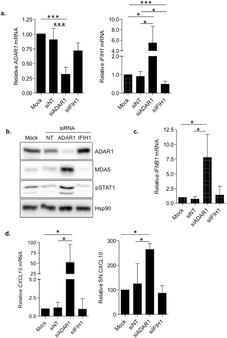
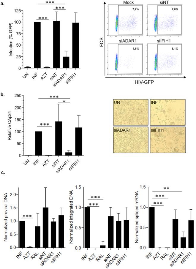
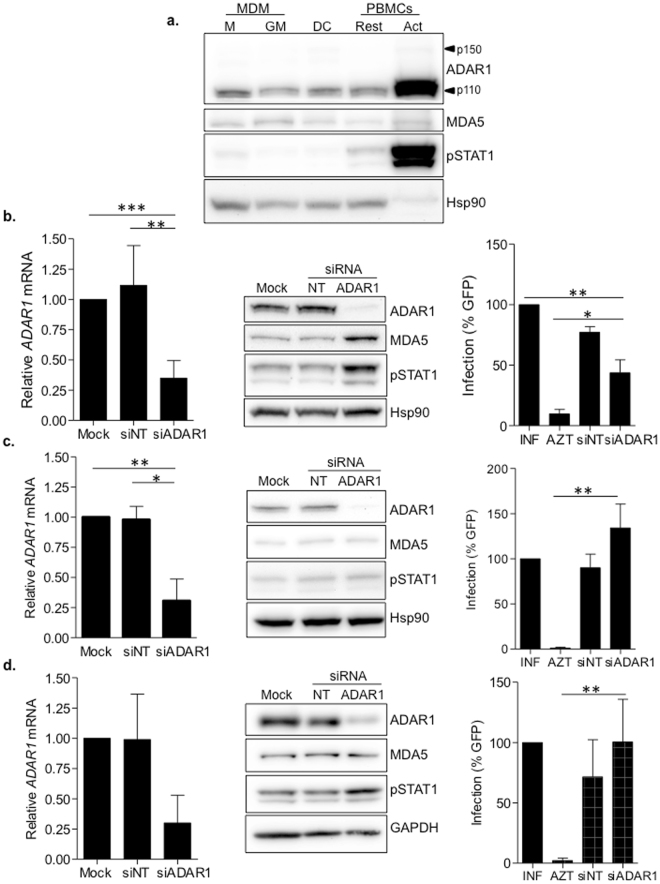
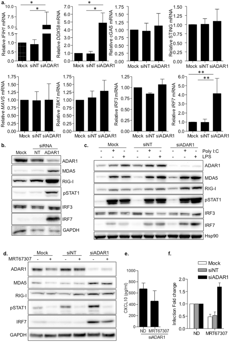
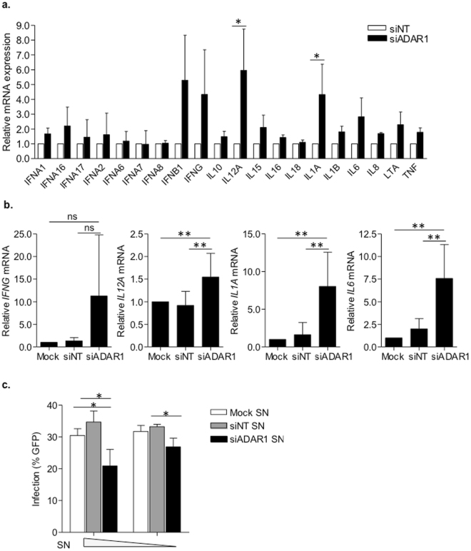
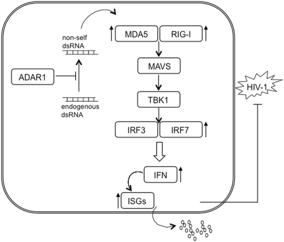
ADAR1-dependent A-to-I editing has recently been recognized as a key process for marking dsRNA as self, therefore, preventing innate immune activation and affecting the development and resolution of immune-mediated diseases and infections. Here, we have determined the role of ADAR1 as a regulator of innate immune activation and modifier of viral susceptibility in primary myeloid and lymphoid cells. We show that ADAR1 knockdown significantly enhanced interferon, cytokine and chemokine production in primary macrophages that function as antiviral paracrine factors, rendering them resistant to HIV-1 infection. ADAR1 knockdown induced deregulation of the RLRs-MAVS signaling pathway, by increasing MDA5, RIG-I, IRF7 and phospho-STAT1 expression, an effect that was partially rescued by pharmacological blockade of the pathway. In summary, our results demonstrate a role of ADAR1 in regulating innate immune function in primary macrophages, suggesting that macrophages may play an essential role in disease associated to ADAR1 dysfunction. We also show that viral inhibition is exclusively dependent on innate immune activation consequence of ADAR1 knockdown, pointing towards ADAR1 as a potential target to boost antiviral immune response.


Conflict of interest statement

The authors declare that they have no competing interests.

Figures

Figure 1
Figure 1
ADAR1 knockdown boosts type I IFN response in primary macrophages. (a) Gene expression of ADAR1 (left panel) and IFIH1 (right panel) knockdown macrophages. Relative mRNA expression of ADAR1 and IFIH1 was measured by quantitative PCR and normalized to GAPDH expression. Data represents mean±SD of 5 different donors and is normalized to mock-transfected M-CSF macrophages. (b) Protein expression in ADAR1 and IFIH1 knockdown macrophages. Western blot of ADAR1, MDA5 and phosphorylation of STAT1 (pSTAT1) in siRNA-treated M-CSF macrophages. MDA5 and pSTAT1 are increased in ADAR1 knockdown macrophages compared to the corresponding non-targeting siRNA (NT). Hsp90 was used as loading control. A representative donor is shown. The figure shows the cropped gels/blots obtained by each protein evaluation. Full-length blots of each tested protein are included in supplementary material. (c) Relative mRNA expression of IFNβ in siRNA-treated macrophages measured by quantitative PCR and normalized to GAPDH expression. IFNB1 gene expression was significantly enhanced in ADAR1 knockdown macrophages. Data represents mean ± SD of 5 different donors and is normalized to mock-transfected M-CSF macrophages. (d) CXCL10 mRNA (left panel) and protein expression in the supernatant (right panel) in siRNA-treated macrophages. Relative mRNA expression of CXCL10 was measured by quantitative PCR and normalized to GAPDH expression. CXCL10 protein in the culture supernatants was measured by ELISA. Data represents mean ± SD of 3 different donors. In all panels, isolated monocytes were transfected with the corresponding siRNA and differentiated to macrophages with M-CSF for 4 days. *p < 0.05; **p < 0.005; ***p < 0.0005.
Figure 2
Figure 2
ADAR1 knockdown blocks HIV-1 transcription in primary macrophages. (a) HIV-1 replication in ADAR1 and IFIH1 knockdown M-CSF macrophages, infected with a VSV-pseudotyped, GFP-expressing HIV-1. Infection was measured 72 h later by flow cytometry. Data represent percentage replication relative to mock-transfected macrophages (left panel). A representative flow cytometry dot plot showing infected macrophages is also depicted (right panel). Mean ± SD of at least 5 different donors performed in triplicate is shown. (b) HIV-1 replication in ADAR1 and IFIH1 knockdown M-CSF macrophages, infected with a full replicative HIV-1 BaL strain. Differentiated macrophages were infected for 7 days and CAp24 production was measured in culture supernatant by ELISA. Mean ± SD of 3 different donors performed in triplicate is shown. (c) Proviral DNA formation (left panel), viral integration (middle panel) and viral transcription (right panel) in ADAR1 and IFIH1 knockdown macrophages. siRNA-treated and subsequently differentiated macrophages were infected with HIV-1 BaL for 16 h (for proviral DNA formation), 20 h (for viral DNA integration) or 40 h (for viral transcription) before DNA or RNA extraction. All determinations were normalized to mock-treated infected macrophages and AZT (3 µM) or raltegravir (RAL; 2 µM), were included as controls. Mean ± SD of at least 3 different donors is shown. In all panels, isolated monocytes were transfected with the corresponding siRNA and differentiated to macrophages with M-CSF for 4 days, prior to infection with HIV-1- *p < 0.05; **p < 0.005; ***p < 0.0005.
Figure 3
Figure 3
ADAR1-mediated regulation of innate immune activation and HIV-1 infection is specific of macrophages. (a) Evaluation of ADAR, MDA5 and pSTAT1 protein expression in different myeloid and lymphoid primary cells. ADAR1 and pSTAT1 expression were increased in activated PBMCs compared to other cell types. M; M-CSF macrophages, GM; GM-CSF macrophages, DC; monocyte derived dendritic cells, Rest; Resting PBMCs and Act, Activated PBMCs. Hsp90 was used as loading control. A representative experiment is shown. (b–d) Evaluation of ADAR1 knockdown in GM-CSF macrophages (b), monocyte-derived dendritic cells (c) and CD4+ T cells (d). Downregulation of ADAR1 mRNA expression by qPCR (left panels), western blot showing protein expression (middle panels) and susceptibility to HIV-1 infection (right panels) are shown. Effective ADAR1 mRNA inhibition was achieved in all cell types (B-D, left panels). Upregulation of MDA5 and pSTAT1 was only observed in ADAR1 knockdown GM-CSF macrophages (b, middle panel), which correlated with inhibition of HIV-1 replication (B, right panel). No significant differences were observed in protein expression or HIV-1 infection in dendritic cells or CD4+ T cells, following ADAR1 knockdown (C and D, middle and right panels respectively). Data from mRNA expression and HIV-1 infection represent the mean ± SD of 3 different donors. A representative western blot is shown in each case. The figure shows the cropped gels/blots obtained by each protein evaluation. Full-length blots of each tested protein are included in supplementary material. *p < 0.05; **p < 0.005.
Figure 4
Figure 4
ADAR1 specifically regulates the RLRs-MAVS signaling pathway. (a) Relative mRNA expression of RLRs (IFIH1 and DDX58), DNA sensors (cGAS and STING), downstream signaling proteins (MAVS, TBK1) and transcription factors (IRF3 and IRF7) in ADAR1 knockdown macrophages. Data represents mean ± SD of at least 4 different donors and is normalized to Mock-transfected macrophages. *p < 0.05; **p < 0.005. (b) Protein expression of RLRs and related proteins in macrophages, showing overexpression of MDA5, RIG-I, pSTAT1 and IRF7 consequence of ADAR1 inhibition. A representative donor is shown. (c) Western blot showing protein expression pattern in transfected macrophages, stimulated or not with LPS (100 ng/ml) or Poly I:C (10 µg/ml) to resemble canonical TLR-mediated activation of type I IFN response. A representative donor is shown. (d) Protein expression in ADAR knockdown macrophages treated with the TBK1 inhibitor MRT67307 (5 µM). Blocking TBK1 function partially restores protein expression phenotype observed in Mock- or siNT-transfected macrophages. A representative donor is shown. (e) CXCL10 protein expression in the supernatant in ADAR1 knockdown macrophages, treated or not with MRT67307 (5 µM). CXCL10 protein in the culture supernatants was measured by ELISA. Data represents mean ± SD of 3 different donors. (f) Change in HIV-1 replication of macrophages treated with the TBK1 inhibitor MRT67307 (5 µM). Fold change of HIV-1 infection in macrophages treated or not with MRT67307. Infection is normalized to the corresponding untreated condition. Data represents mean ± SD of 3 different donors performed in triplicate. *p < 0.05; **p < 0.005. The figure shows the cropped gels/blots obtained by each protein evaluation. Full-length blots of each tested protein are included in supplementary material.
Figure 5
Figure 5
ADAR1 knockdown macrophages present a pro-inflammatory phenotype that inhibits HIV-1 replication of bystander cells. (a) Cytokine expression profile of ADAR1 knockdown macorphages. Relative mRNA expression of the different cytokines normalized to siNT-transfected macrophages is depicted. Data represents mean ± SD of 3 different donors. *p < 0.05. (b) Relative mRNA expression of cytokines identified as upregulated in ADAR1 knockdown macrophages (IFNG, IL12A, IL1A and IL6). Data represents mean ± SD of 6 independent donors and is normalized to mock-transfected macrophages. ns; non-significant, **p < 0.005. (c) Antiviral activity of culture supernatants from ADAR1 knockdown macrophages. Percentage infection of untreated macrophages incubated with different concentrations of culture supernatants from mock- (white bars), siNT- (grey bars) and siADAR1- (black bars) treated macrophages. SN; supernatant, *p < 0.05.
Figure 6
Figure 6
Representation of signaling cascade regulated by ADAR1 in MDM. ADAR1 modulates the recognition of non-self dsRNA in the cytoplasm; consequently, sensing of non-self dsRNA induces expression of RNA sensors, RIG-I and MDA5, through downstream effectors, MAVS and TBK1, and transcription factors IRF3 and IRF7. Leading to induction of an innate immune response characterized by increase expression of type I IFN and ISG and subsequent over production of cytokines and chemokines able to block HIV-1.

Similar articles

Cited by

References

    1. George, C. X., John, L. & Samuel, C. E. An RNA editor, adenosine deaminase acting on double-stranded RNA (ADAR1). Journal of interferon & cytokine research: the official journal of the International Society for Interferon and Cytokine Research 34, 437–446, 10.1089/jir.2014.0001 (2014). - PMC - PubMed
    1. Nishikura K. A-to-I editing of coding and non-coding RNAs by ADARs. Nature reviews. Molecular cell biology. 2016;17:83–96. doi: 10.1038/nrm.2015.4. - DOI - PMC - PubMed
    1. Song, C., Sakurai, M., Shiromoto, Y. & Nishikura, K. Functions of the RNA Editing Enzyme ADAR1 and Their Relevance to Human Diseases. Genes7, 10.3390/genes7120129 (2016). - PMC - PubMed
    1. Wang, Q., Li, X., Qi, R. & Billiar, T. RNA Editing, ADAR1, and the Innate Immune Response. Genes8, 10.3390/genes8010041 (2017). - PMC - PubMed
    1. Liddicoat BJ, et al. RNA editing by ADAR1 prevents MDA5 sensing of endogenous dsRNA as nonself. Science. 2015;349:1115–1120. doi: 10.1126/science.aac7049. - DOI - PMC - PubMed

Publication types

MeSH terms