

Screening of differentially expressed proteins in psoriasis vulgaris by two-dimensional gel electrophoresis and mass spectrometry
- PMID: 29042920
- PMCID: PMC5639297
- DOI: 10.3892/etm.2017.5012
Screening of differentially expressed proteins in psoriasis vulgaris by two-dimensional gel electrophoresis and mass spectrometry
Abstract
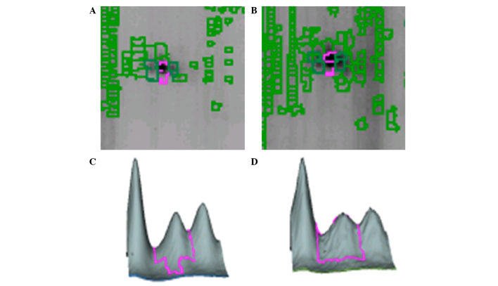
The aim of the present study was to elucidate differentially expressed proteins in lesional tissues of psoriasis vulgaris (PV) and normal tissues. Lesional skin tissues were collected from PV patients, along with normal skin tissues from healthy individuals. The protein content of the samples was extracted and then separated by two-dimensional gel electrophoresis (2-DGE). Any proteins that were differentially expressed in the lesional skin of PV patients compared with the healthy controls were analyzed by mass spectrometry and bioinformatics. In the stratum corneum and dermis of PV patients, the total number of proteins identified by 2-DGE was 1,969±21 and 1,928±49, respectively. Of these, 30 proteins were differentially expressed in the PV patients, of which 14 were identified as: Type 1 keratin cytoskeleton proteins (including K1C10, K1C14, K1C15 and K1C16); the type 2 keratin cytoskeleton protein, K2C1; actin-associated proteins (including ARP3, ACTA and ACTBM); prohibitin; heat shock proteins (HSPB1 and CH60); centrosome protein, CP135; and membrane associated proteins (including ANXA4 and ANXA5). The differential expression of protein between PV lesions and normal tissue can be considered as pathological biomarker. Elucidating the abnormal regulation of these proteins can provide mechanism of the development of PV and may contribute to significant approaches for PV treatments.
Keywords: differentially expressed proteins; mass spectrometry; psoriasis vulgaris; two-dimensional gel electrophoresis.
Figures


Similar articles
-
Differences in expression of serum protein in patients with psoriasis vulgaris and blood heat syndrome and healthy volunteers.Cell Mol Biol (Noisy-le-grand). 2017 Aug 30;63(8):38-41. doi: 10.14715/cmb/2017.63.8.9. Cell Mol Biol (Noisy-le-grand). 2017. PMID: 28886312
-
Quantitative analysis of differentially expressed proteins in psoriasis vulgaris using tandem mass tags and parallel reaction monitoring.Clin Proteomics. 2020 Aug 12;17:30. doi: 10.1186/s12014-020-09293-8. eCollection 2020. Clin Proteomics. 2020. PMID: 32817748 Free PMC article.
-
Proteomic analysis of psoriatic skin tissue for identification of differentially expressed proteins: up-regulation of GSTP1, SFN and PRDX2 in psoriatic skin.Int J Mol Med. 2011 Nov;28(5):785-92. doi: 10.3892/ijmm.2011.757. Epub 2011 Jul 25. Int J Mol Med. 2011. PMID: 21805023
-
Proteome analysis of skin distinguishes acute guttate from chronic plaque psoriasis.J Invest Dermatol. 2005 Jan;124(1):63-9. doi: 10.1111/j.0022-202X.2004.23501.x. J Invest Dermatol. 2005. PMID: 15654954
-
Novel findings from determination of common expressed plasma exosomal microRNAs in patients with psoriatic arthritis, psoriasis vulgaris, rheumatoid arthritis, and gouty arthritis.Discov Med. 2019 Jul;28(151):47-68. Discov Med. 2019. PMID: 31465725
Cited by
-
Changes in Proteome of Fibroblasts Isolated from Psoriatic Skin Lesions.Int J Mol Sci. 2020 Jul 28;21(15):5363. doi: 10.3390/ijms21155363. Int J Mol Sci. 2020. PMID: 32731552 Free PMC article.
References
-
- Barker JN. Psoriasis as a T cell-mediated autoimmune disease. Hosp Med. 1998;59:530–533. - PubMed
-
- Shakery K, Reich K. Psoriasis-clinical picture and current therapy. Med Monatsschr Pharm. 2009;32:335–344. quiz 345–346. (In German) - PubMed
-
- Reich A, Szepietowski J. Genetic and immunological aspects of the pathogenesis of psoriasis. Wiad Lek. 2007;60:270–276. (In Polish) - PubMed
LinkOut - more resources
Full Text Sources
Other Literature Sources
Research Materials
Miscellaneous