

Endoscopy in the treatment of slit ventricle syndrome
- PMID: 29042922
- PMCID: PMC5639315
- DOI: 10.3892/etm.2017.4973
Endoscopy in the treatment of slit ventricle syndrome
Abstract
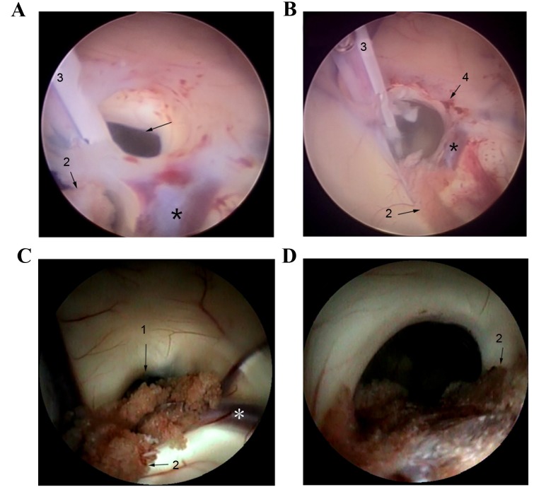
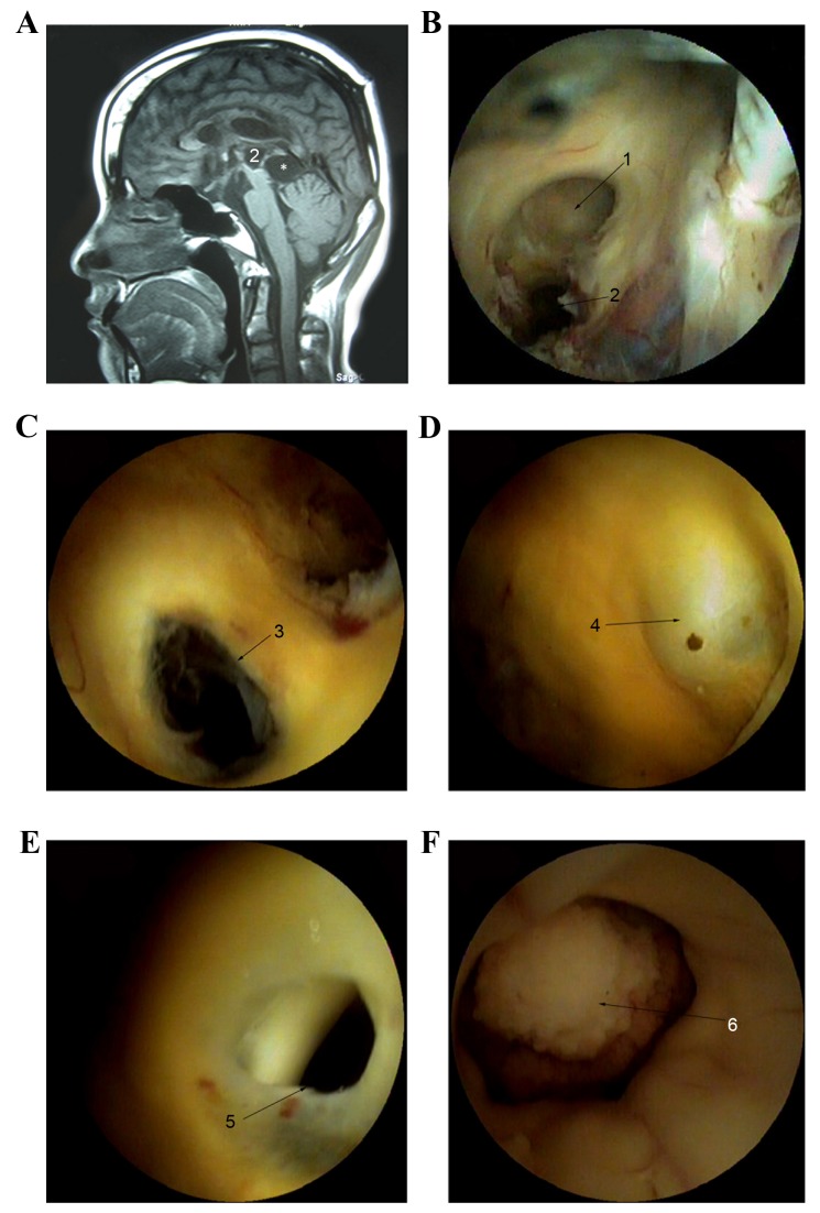
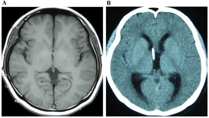
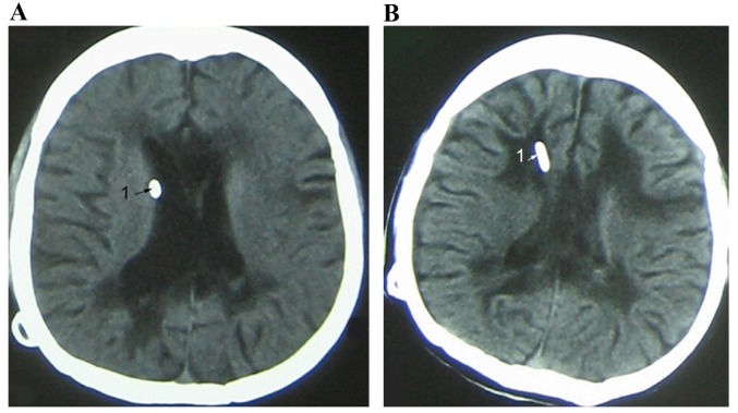
The present study aimed to investigate the efficacy of endoscopy in the treatment of post-shunt placement for slit ventricle syndrome (SVS). Endoscopic surgery was performed on 18 patients with SVS between October 2004 and December 2012. Sex, age, causes of the hydrocephalus, ventricular size and imaging data were collected and analyzed. All patients were divided into two groups according to ventricular size and underwent endoscopic surgeries, including endoscopic third ventriculostomy (ETV), endoscopic aqueductoplasty and cystocisternostomy. All treated patients were observed postoperatively for a period of 2 to 3 weeks, and outpatient follow-up was subsequently scheduled for >12 months. Clinical results, including catheter adherence, shunt removal and complications, were analyzed during the follow-up period. The success rate of endoscopic surgery was indicated to be 82.7%. Syndromes caused by aqueductal stenosis in 15 patients who underwent ETV were relieved; however, syndromes in the 3 patients with cerebral cysticercosis, suprasellar arachnoid cysts, pinea larea glioma and communicating hydrocephalus, respectively, were not relieved and underwent shunt placement again. Brain parenchyma, choroid plexus and ependymal tissue were the predominant causes for catheter obstruction and the obstruction rate was indicated to be 77.8% (14/18). Complications, such as pseudobulbar paralysis, infection and intraventricular hemorrhage arose in 3 patients. The present study indicates that endoscopic treatments are effective and ETV may be considered as a recommended option in the treatment of post-shunt placement SVS in hydrocephalus patients.
Keywords: endoscopy; hydrocephalus; slit ventricle syndrome; ventriculoperitoneal shunt.
Figures
Similar articles
-
Comparison of hydrocephalus metrics between infants successfully treated with endoscopic third ventriculostomy with choroid plexus cauterization and those treated with a ventriculoperitoneal shunt: a multicenter matched-cohort analysis.J Neurosurg Pediatr. 2018 Apr;21(4):339-345. doi: 10.3171/2017.10.PEDS17421. Epub 2018 Feb 2. J Neurosurg Pediatr. 2018. PMID: 29393809
-
Endoscopic third ventriculostomy and choroid plexus cauterization in infant hydrocephalus: a prospective study by the Hydrocephalus Clinical Research Network.J Neurosurg Pediatr. 2018 Mar;21(3):214-223. doi: 10.3171/2017.8.PEDS17217. Epub 2017 Dec 15. J Neurosurg Pediatr. 2018. PMID: 29243972
-
Endoscopic third ventriculostomy and choroid plexus cauterization with a rigid neuroendoscope in infants with hydrocephalus.J Neurosurg Pediatr. 2016 Feb;17(2):163-173. doi: 10.3171/2015.5.PEDS14692. Epub 2015 Oct 30. J Neurosurg Pediatr. 2016. PMID: 26517057
-
Endoscopic third ventriculostomy for idiopathic aqueductal stenosis.World Neurosurg. 2013 Feb;79(2 Suppl):S21.e13-20. doi: 10.1016/j.wneu.2012.02.007. Epub 2012 Feb 10. World Neurosurg. 2013. PMID: 22381825 Review.
-
Endoscopic re-opening of third ventriculostomy: Case series and review of literature.Clin Neurol Neurosurg. 2016 Jun;145:58-63. doi: 10.1016/j.clineuro.2016.04.007. Epub 2016 Apr 11. Clin Neurol Neurosurg. 2016. PMID: 27088221 Review.
Cited by
-
Slit ventricle syndrome: Historical considerations, diagnosis, pathophysiology, and treatment review.Brain Circ. 2021 Aug 27;7(3):167-177. doi: 10.4103/bc.bc_29_21. eCollection 2021 Jul-Sep. Brain Circ. 2021. PMID: 34667900 Free PMC article. Review.
-
Image quality and related outcomes of the ShuntScope for catheter implantation in pediatric hydrocephalus-experience of 65 procedures.Childs Nerv Syst. 2023 Mar;39(3):721-732. doi: 10.1007/s00381-022-05776-1. Epub 2022 Dec 2. Childs Nerv Syst. 2023. PMID: 36459211 Free PMC article.
-
Suprasellar arachnoid cysts: systematic analysis of 247 cases with long-term follow-up.Neurosurg Rev. 2021 Oct;44(5):2755-2765. doi: 10.1007/s10143-020-01455-4. Epub 2021 Jan 7. Neurosurg Rev. 2021. PMID: 33409764
References
LinkOut - more resources
Full Text Sources
Other Literature Sources