

Delivery of biotinylated IGF-1 with biotinylated self-assembling peptides combined with bone marrow stem cell transplantation promotes cell therapy for myocardial infarction
- PMID: 29042931
- PMCID: PMC5639271
- DOI: 10.3892/etm.2017.4982
Delivery of biotinylated IGF-1 with biotinylated self-assembling peptides combined with bone marrow stem cell transplantation promotes cell therapy for myocardial infarction
Abstract
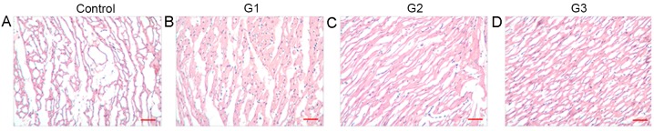
Cell therapy is a promising approach for cardiac repair. The aim of the present study was to determine the feasibility of using biotinylated insulin-like growth factor 1 (IGF-1) with biotinylated self-assembling peptides (tethered IGF-1) combined with bone marrow stem cells (BMSCs) transplantation for the treatment of heart failure. Tethered IGF-1 was synthesized and its effect on H9c2 cells was analyzed. Reverse transcription-quantitative polymerase chain reaction and western blot assays demonstrated that tethered IGF-1 did not significantly affect the expression and phosphorylation of AKT, whereas it significantly increased the expression of cardiac troponin T (P<0.01). A rabbit myocardial infarction model was constructed and rabbits were divided into four groups: Control group (no treatment), group 1 (G1; BMSC transplantation), group 2 (G2; BMSCs + non-biotinylated IGF-1) and group 3 (G3; BMSCs + tethered IGF-1). At 4 weeks after modeling, cardiac tissues were obtained for analysis. In the control group, myocardial fibers were disordered, a large number of inflammatory cells infiltrated the cardiac tissues, and apoptosis occurred in ~50% of cells. However, in G1, G2 and G3, muscle cells were well ordered, and a lesser degree of myocardial degeneration and inflammatory cell infiltration was observed. Compared with the control group, the apoptosis rates of myocardial cells in G1-G3 were significantly decreased (P<0.01). Furthermore, compared with G1 and G2, tissue morphology was improved in G3and the number of apoptotic myocardial cells was significantly decreased (P<0.01). These results suggest that treatment with tethered IGF-1 + BMSCs significantly suppresses cell apoptosis and induces the expression of cardiac maturation proteins. These findings provide a novel insight into how the delivery of tethered IGF-1 with BMSCs could potentially enhance the prognosis of patients with heart failure treatment.
Keywords: bone marrow stem cell; cardiac failure; insulin-like growth factor 1; self-assembling peptides.
Figures


Similar articles
-
HGF and IGF-1 promote protective effects of allogeneic BMSC transplantation in rabbit model of acute myocardial infarction.Cell Prolif. 2015 Dec;48(6):661-70. doi: 10.1111/cpr.12219. Epub 2015 Oct 15. Cell Prolif. 2015. PMID: 26466964 Free PMC article.
-
Local myocardial insulin-like growth factor 1 (IGF-1) delivery with biotinylated peptide nanofibers improves cell therapy for myocardial infarction.Proc Natl Acad Sci U S A. 2006 May 23;103(21):8155-60. doi: 10.1073/pnas.0602877103. Epub 2006 May 12. Proc Natl Acad Sci U S A. 2006. PMID: 16698918 Free PMC article.
-
Hepatocyte growth factor combined with insulin like growth factor-1 improves expression of GATA-4 in mesenchymal stem cells cocultured with cardiomyocytes.Chin Med J (Engl). 2008 Feb 20;121(4):336-40. Chin Med J (Engl). 2008. PMID: 18304467
-
[EXPERIMENTAL STUDY ON LENTIVIRUS-MEDIATED MULTI-GENES CO-TRANSFECTION IN BONE MARROW MESENCHYMAL STEM CELLS FOR TREATMENT OF KNEE OSTEOARTHRITIS IN CYNOMOLGUS MONKEY].Zhongguo Xiu Fu Chong Jian Wai Ke Za Zhi. 2016 Sep 8;30(9):1153-1159. doi: 10.7507/1002-1892.20160235. Zhongguo Xiu Fu Chong Jian Wai Ke Za Zhi. 2016. PMID: 29786374 Chinese.
-
Bone marrow stromal cell transplantation combined with angiotensin-converting enzyme inhibitor treatment in rat with acute myocardial infarction and the role of insulin-like growth factor-1.Cytotherapy. 2012 May;14(5):563-9. doi: 10.3109/14653249.2011.651531. Epub 2012 Jan 25. Cytotherapy. 2012. PMID: 22277013
Cited by
-
Toward Regeneration of the Heart: Bioengineering Strategies for Immunomodulation.Front Cardiovasc Med. 2019 Mar 21;6:26. doi: 10.3389/fcvm.2019.00026. eCollection 2019. Front Cardiovasc Med. 2019. PMID: 30949485 Free PMC article. Review.
-
Analyzing Impetus of Regenerative Cellular Therapeutics in Myocardial Infarction.J Clin Med. 2020 Apr 28;9(5):1277. doi: 10.3390/jcm9051277. J Clin Med. 2020. PMID: 32354170 Free PMC article. Review.
References
-
- Le HH, El-Khatib C, Mombled M, Guitarian F, Al-Gobari M, Fall M, Janiaud P, Marchant I, Cucherat M, Bejan-Angoulvant T, Gueyffier F. Impact of aldosterone antagonists on sudden cardiac death prevention in heart failure and post-myocardial infarction patients: A systematic review and meta-analysis of randomized controlled trials. PLoS One. 2016;11:e0145958. doi: 10.1371/journal.pone.0145958. - DOI - PMC - PubMed
-
- Nygren JM, Jovinge S, Breitbach M, Säwén P, Röll W, Hescheler J, Taneera J, Fleischmann BK, Jacobsen SE. Bone marrow-derived hematopoietic cells generate cardiomyocytes at a low frequency through cell fusion, but not transdifferentiation. Nat Med. 2004;10:494–501. doi: 10.1038/nm1040. - DOI - PubMed
LinkOut - more resources
Full Text Sources
Other Literature Sources
Miscellaneous