

Aphasic status epilepticus as the sole symptom of epilepsy: A case report and literature review
- PMID: 29042939
- PMCID: PMC5639272
- DOI: 10.3892/etm.2017.4979
Aphasic status epilepticus as the sole symptom of epilepsy: A case report and literature review
Abstract
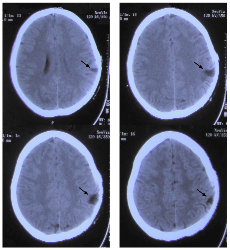
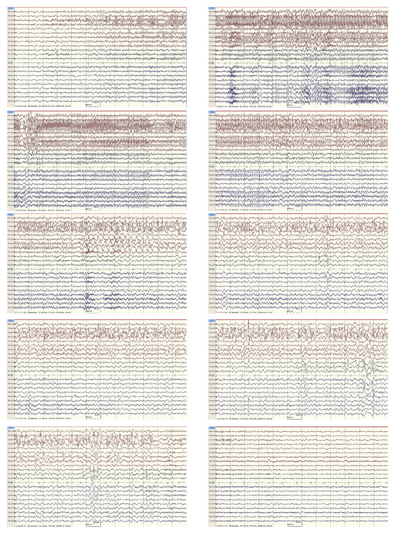
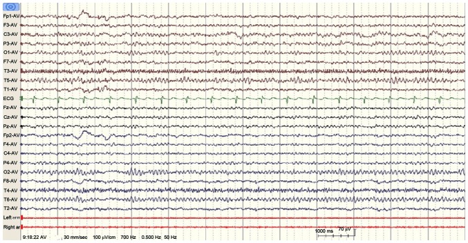
Aphasia is a common symptom encountered by neurologists. However, the presence of aphasia as the sole manifestation of partial status epilepticus is rare. The present study reports a case of aphasic status epilepticus in a 27-year-old right-handed female who presented after the abrupt onset of aphasia, which had persisted for 1.5 days. The patient's medical history included head trauma followed by a craniectomy and cranioplasty. Computed tomography scans revealed a lesion in the patient's left parietal lobe, and an electroencephalogram showed a spike and slow wave pattern in the left hemisphere of the brain during aphasia. The patient's condition improved after the oral administration of oxcarbazepine daily. In the present study it was observed that EEGs were a simple method to diagnose aphasic seizures and therefore EEG recordings should be performed in all cases of unexplained aphasia. In addition, the present study reviewed previously reported cases of aphasic status epilepticus.
Keywords: EEG; aphasia status epilepticus; epilepsy.
Figures
Similar articles
-
Prolonged ictal aphasia: a diagnosis to consider.J Clin Neurosci. 2012 Nov;19(11):1605-6. doi: 10.1016/j.jocn.2012.04.004. Epub 2012 Aug 25. J Clin Neurosci. 2012. PMID: 22925412
-
Aphasic status epilepticus in a tertiary referral center in Turkey: Clinical features, etiology, and outcome.Epilepsy Res. 2020 Nov;167:106479. doi: 10.1016/j.eplepsyres.2020.106479. Epub 2020 Sep 29. Epilepsy Res. 2020. PMID: 33038720
-
Aphasic status epilepticus preceding tumefactive left hemisphere lesion in anti-MOG antibody associated disease.Mult Scler Relat Disord. 2019 Jan;27:91-94. doi: 10.1016/j.msard.2018.10.012. Epub 2018 Oct 15. Mult Scler Relat Disord. 2019. PMID: 30347340
-
De novo aphasic status epilepticus.Epilepsia. 1997 Aug;38(8):945-9. doi: 10.1111/j.1528-1157.1997.tb01262.x. Epilepsia. 1997. PMID: 9579898 Review.
-
Nonconvulsive status epilepticus presenting as a subacute progressive aphasia.Seizure. 2002 Oct;11(7):449-54. doi: 10.1053/seiz.2002.0678. Seizure. 2002. PMID: 12237073 Review.
Cited by
-
Aphasic Status Epilepticus Associated with Alzheimer's Disease: Clinical and Electrographic Characteristics.J Epilepsy Res. 2023 Dec 31;13(2):55-58. doi: 10.14581/jer.23009. eCollection 2023 Dec. J Epilepsy Res. 2023. PMID: 38223360 Free PMC article.
-
Verbal and memory deficits caused by aphasic status epilepticus after resection of a left temporal lobe glioma.Surg Neurol Int. 2021 Dec 14;12:614. doi: 10.25259/SNI_1120_2021. eCollection 2021. Surg Neurol Int. 2021. PMID: 34992930 Free PMC article.
-
Follow-Up in Aphasia Caused by Acute Stroke in a Prospective, Randomized, Clinical, and Experimental Controlled Noninvasive Study With an iPad-Based App (Neolexon®): Study Protocol of the Lexi Study.Front Neurol. 2020 Apr 30;11:294. doi: 10.3389/fneur.2020.00294. eCollection 2020. Front Neurol. 2020. PMID: 32425873 Free PMC article.
-
Three Cases of Aphasic Status Epilepticus: Clinical and Electrographic Characteristics.Clin Med Insights Case Rep. 2021 Apr 10;14:11795476211009241. doi: 10.1177/11795476211009241. eCollection 2021. Clin Med Insights Case Rep. 2021. PMID: 33953631 Free PMC article.
-
Aphasia with No Apparent Paralysis in Progressive Stroke of the Anterior Choroidal Artery.Intern Med. 2023 Apr 1;62(7):1059-1062. doi: 10.2169/internalmedicine.0009-22. Epub 2022 Aug 30. Intern Med. 2023. PMID: 36047127 Free PMC article.
References
LinkOut - more resources
Full Text Sources
Other Literature Sources