

Diagnosis and treatment of pulmonary mucormycosis: A case report
- PMID: 29042980
- PMCID: PMC5639346
- DOI: 10.3892/etm.2017.4986
Diagnosis and treatment of pulmonary mucormycosis: A case report
Abstract
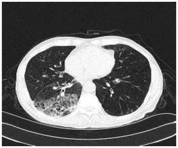
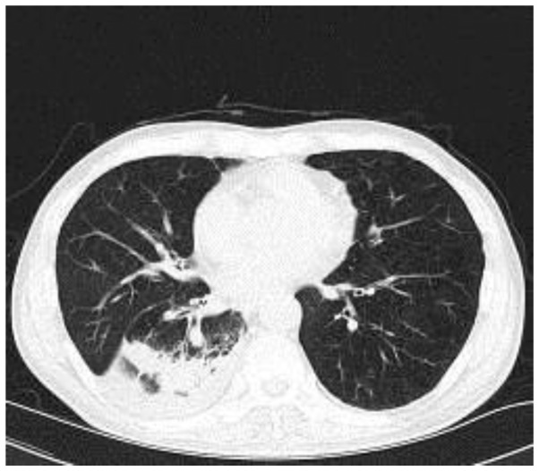
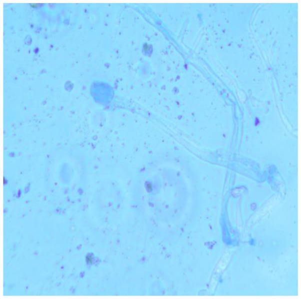
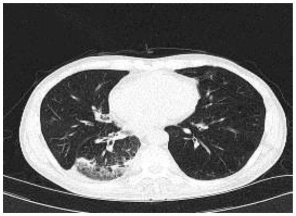
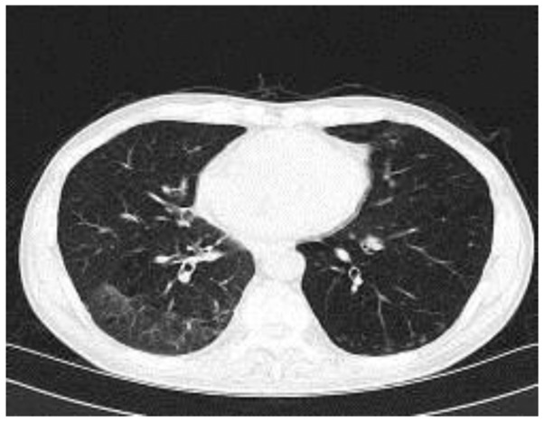
Pulmonary mucormycosis, a relatively rare pulmonary fungal disease, is difficult to diagnose and lacks effective treatment. The present study reports the case of a 64-year-old patient who was treated successfully for pulmonary mucormycosis in Xuan Wu Hospital. The patient presented with low-grade fever and a productive cough that persisted for 1 month with no evident cause, and also suffered from diabetes mellitus. Prior antibiotic treatment with levofloxacin had been ineffective. Culture of mucus obtained by bronchoscopy resulted in a diagnosis of pulmonary mucormycosis. The patient received a cumulative dose of 1,355 mg amphotericin B over 2 months and presented a full recovery.
Keywords: diagnosis; pulmonary mucormycosis; treatment.
Figures
Similar articles
-
[Pulmonary mucormycosis: report of 5 cases and review of 46 cases reported in China].Zhonghua Jie He He Hu Xi Za Zhi. 2013 Aug;36(8):572-6. Zhonghua Jie He He Hu Xi Za Zhi. 2013. PMID: 24252732 Chinese.
-
Pulmonary Mucormycosis: A Case Report of a Rare Infection with Potential Diagnostic Problems.Case Rep Pathol. 2020 Jan 6;2020:5845394. doi: 10.1155/2020/5845394. eCollection 2020. Case Rep Pathol. 2020. PMID: 31970007 Free PMC article.
-
Invasive pulmonary mucormycosis: rare presentation with pulmonary eosinophilia.BMC Pulm Med. 2017 Apr 28;17(1):76. doi: 10.1186/s12890-017-0419-1. BMC Pulm Med. 2017. PMID: 28454572 Free PMC article.
-
Report of 12 cases with tracheobronchial mucormycosis and a review.Clin Respir J. 2018 Apr;12(4):1651-1660. doi: 10.1111/crj.12724. Epub 2018 Feb 19. Clin Respir J. 2018. PMID: 29028140 Review.
-
Pulmonary mucormycosis in a diabetic patient.Ann Med Interne (Paris). 2000 Dec;151(8):669-72. Ann Med Interne (Paris). 2000. PMID: 11173713 Review.
Cited by
-
Breaking the mold: A case of pulmonary mucormycosis treated with isavuconazole.Med Mycol Case Rep. 2018 Dec 1;23:34-36. doi: 10.1016/j.mmcr.2018.11.004. eCollection 2019 Mar. Med Mycol Case Rep. 2018. PMID: 30560048 Free PMC article.
-
Oral Isavuconazole Combined with Nebulized Inhalation and Bronchoscopic Administration of Amphotericin B for the Treatment of Pulmonary Mucormycosis: A Case Report and Literature Review.J Fungi (Basel). 2024 May 29;10(6):388. doi: 10.3390/jof10060388. J Fungi (Basel). 2024. PMID: 38921374 Free PMC article.
-
Pulmonary mucormycosis in immunocompetent hosts diagnosed by bronchioalveolar lavage.BMJ Case Rep. 2021 Apr 12;14(4):e240180. doi: 10.1136/bcr-2020-240180. BMJ Case Rep. 2021. PMID: 33846183 Free PMC article.
-
Pulmonary mucormycosis mimicking lung tumour in an uncontrolled diabetic patient.Respirol Case Rep. 2022 Feb 23;10(3):e0917. doi: 10.1002/rcr2.917. eCollection 2022 Mar. Respirol Case Rep. 2022. PMID: 35228889 Free PMC article.
-
Case series of invasive lung infections by Aspergillus species and zygomycosis among post COVID-19 and post-transplant individuals.J Family Med Prim Care. 2022 Nov;11(11):7469-7475. doi: 10.4103/jfmpc.jfmpc_1066_22. Epub 2022 Dec 16. J Family Med Prim Care. 2022. PMID: 36993027 Free PMC article.
References
-
- Liu YN, She DY, Sun TY, Tong ZH, He B, Xiao Y, He LX, Qu JM, Liu XQ, Li ER, et al. A multicenter retrospective study of pulmonary mycosis clinically proven from 1998–2007. Zhonghua Jie He He Hu Xi Za Zhi. 2011;34:86–90. (In Chinese) - PubMed
-
- Yang Y, Fang B, Xu X, Fang F, Pan M, Zhong X, Sun T. Pulmonary mucormycosis: report of 5 cases and review of 46 cases reported in China. Chin J Tuberc Respir Dis. 2013;36:572–576. (In Chinese) - PubMed
LinkOut - more resources
Full Text Sources
Other Literature Sources