. 2017 Sep;50(3):230-238.
doi: 10.5115/acb.2017.50.3.230. Epub 2017 Sep 20.

The distribution of calbindin-D28k, parvalbumin, and calretinin immunoreactivity in the inferior colliculus of circling mouse

Affiliations

The distribution of calbindin-D28k, parvalbumin, and calretinin immunoreactivity in the inferior colliculus of circling mouse

Jin-Koo Lee et al. Anat Cell Biol. 2017 Sep.

Abstract

The circling mice with tmie gene mutation are known as an animal deafness model, which showed hyperactive circling movement. Recently, the reinvestigation of circling mouse was performed to check the inner ear pathology as a main lesion of early hearing loss. In this trial, the inner ear organs were not so damaged to cause the hearing deficit of circling (cir/cir) mouse at 18 postnatal day (P18) though auditory brainstem response data indicated hearing loss of cir/cir mice at P18. Thus, another mechanism may be correlated with the early hearing loss of cir/cir mice at P18. Hearing loss in the early life can disrupt the ascending and descending information to inferior colliculus (IC) as integration site. There were many reports that hearing loss could result in the changes in Ca2+ concentration by either cochlear ablation or genetic defect. However, little was known to be reported about the correlation between the pathology of IC and Ca2+ changes in circling mice. Therefore, the present study investigated the distribution of calcium-binding proteins (CaBPs), calbindin-D28k, parvalbumin, and calretinin immunoreactivity (IR) in the IC to compare among wild-type (+/+), heterozygous (+/cir), and homozygous (cir/cir) mice by immunohistochemistry. The decreases of CaBPs IR in cir/cir were statistically significant in the neurons as well as neuropil of IC. Thus, this study proposed overall distributional alteration of CaBPs IR in the IC caused by early hearing defect and might be helpful to elucidate the pathology of central auditory disorder related with Ca2+ metabolism.

Keywords: Calbindin-D28k; Calretinin; Circling mice; Inferior colliculus; Parvalbumins.


Figures

Fig. 1
Fig. 1. Distribution of calbindin-D28k (CB) immunoreactivity (IR) in coronal sections through the inferior colliculus (IC) of postnatal day 16 wild-type (+/+) (A, D, G), heterozygous (+/cir) (B, E, H), and homozygous (cir/cir) (C, F, I) mice. CB IR was noted to be drastically decreased in both +/cir and cir/cir as compared with +/+ in the whole IC region. Prominent loss of staining was noted in the cells (arrows) of both outer dorsal cortex of IC (DC) and inner DC of cir/cir (D–I). Oval to round in shape cells (arrows) with very short proximal dendritic staining along with immunoreactive fibers were noted in the central nucleus of IC region (J–L). Loss of such cells and fibers were noted in cir/cir. The lateral nucleus of IC region consisted of oblong, stellate and oval soma (arrows) which were darkly stained in +/+ but was very faint in +/cir and cir/cir (M–O). Scale bars=100 µm (A–C), 50 µm (D–O).
Fig. 2
Fig. 2. Distribution of parvalbumin (PV) immunoreactivity (IR) in coronal sections through the inferior colliculus (IC) of postnatal day 16 wild-type (+/+) (A, D, G), heterozygous (+/cir) (B, E, H), and homozygous (cir/cir) (C, F, I) mice. PV IR was distributed with varying intensity which was slightly lower in cir/cir and the labeling intensity was noted to be highest in the dorsolateral part of IC. Oval to multipolar cells (arrows) were stained in both the outer dorsal cortex of IC (DC) and inner DC region (D–I). Numerous PV immunoreactive cells (arrows) were also present in the central nucleus of IC region of all the three types which appeared to be oval, spindle and multipolar in shape (J–L). Lateral nucleus of IC region consisted of cells (arrows) displaying proximal dendritic staining and immunoreactive fibers as well (M–O). Scale bars=100 µm (A–C), 50 µm (D–O).
Fig. 3
Fig. 3. Distribution of calretinin (CR) immunoreactivity (IR) in coronal sections through the inferior colliculus (IC) of postnatal day 16 wild-type (+/+) (A, D, G), heterozygous (+/cir) (B, E, H), and homozygous (cir/cir) (C, F, I) mice. Highly stained cells were noted in dorsal cortex of IC (DC) and lateral nucleus of IC (LN) region but were very less in central nucleus of IC (ICC) of all the three types with varying intensity. Flattened cells (arrows) with dendrites sprouting out was observed in the outer DC (D–F) while the inner DC consisted of triangular, rounded or occasionally round shaped cells (arrows) (G–I). Diffuse background staining of neuropil prominent in +/+ was lacking in cir/cir. lightly stained cells (arrows) and diffuse neuropil staining was noted in ICC which decreased prominently in cir/cir (J–L). CR immunoreactive lemniscal tract fibers were also present. Numerous CR positive cells (arrows) with dendrites running parallel to the surface of IC along with immunoreactive fibers were noted in the LN region (M–O). CR IR in cells, fibers, and neuropil appeared to be prominently decrease in all the regions of IC of cir/cir as compared with +/+. Scale bars=100 µm (A–C), 50 µm (D–O).
Fig. 4
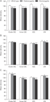
Fig. 4. Image analysis of relative density of calbindin-D28k (A), parvalbumin (B), and calretinin (C) immunoreactivity (IR) changes in the inferior colliculus (IC) of postnatal day 16 wild-type (+/+), heterozygous (+/cir), and homozygous (cir/cir) mice. Decrease in the IR was noted in cir/cir in the various different regions of IC as compared with +/+. The data shown are the mean±SD obtained from three different experiments. DC, dorsal cortex of IC; ICC, central nucleus of IC; LN, lateral nucleus of IC. **P<0.01, ***P<0.001, compared with wild-type.

Similar articles

Cited by

References

    1. Shin MJ, Lee JH, Yu DH, Kim BS, Kim HJ, Kim SH, Kim MO, Park C, Hyun BH, Lee S, So HS, Park R, Ryoo ZY. Ectopic expression of tmie transgene induces various recovery levels of behavior and hearing ability in the circling mouse. Biochem Biophys Res Commun. 2008;374:17–21. - PubMed
    1. Cho KI, Lee JW, Kim KS, Lee EJ, Suh JG, Lee HJ, Kim HT, Hong SH, Chung WH, Chang KT, Hyun BH, Oh YS, Ryoo ZY. Fine mapping of the circling (cir) gene on the distal portion of mouse chromosome 9. Comp Med. 2003;53:642–648. - PubMed
    1. Chung WH, Kim KR, Cho YS, Cho DY, Woo JH, Ryoo ZY, Cho KI, Hong SH. Cochlear pathology of the circling mouse: a new mouse model of DFNB6. Acta Otolaryngol. 2007;127:244–251. - PubMed
    1. Lee Y, Chang SY, Jung JY, Ahn SC. Reinvestigation of cochlear pathology in circling mice. Neurosci Lett. 2015;594:30–35. - PubMed
    1. Illing RB. Activity-dependent plasticity in the adult auditory brainstem. Audiol Neurootol. 2001;6:319–345. - PubMed

LinkOut - more resources