Review
. 2017 Oct;47(11):1431-1440.
doi: 10.1007/s00247-017-3891-0. Epub 2017 Sep 21.

Computed tomography in children with community-acquired pneumonia

Affiliations
Review

Computed tomography in children with community-acquired pneumonia

Savvas Andronikou et al. Pediatr Radiol. 2017 Oct.

Abstract

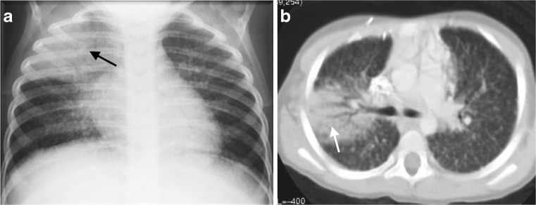
Diagnostic imaging plays a significant role in both the diagnosis and treatment of complications of pneumonia in children and chest radiography is the imaging modality of choice. Computed tomography (CT) on the other hand, is not currently a first-line imaging tool for children with suspected uncomplicated community-acquired pneumonia and is largely reserved for when complications of pneumonia are suspected or there is difficulty in differentiating pneumonia from other pathology. This review outlines the situations where CT needs to be considered in children with pneumonia, describes the imaging features of the parenchymal and pleural complications of pneumonia, discusses how CT may have a wider role in developing countries where human immunodeficiency virus (HIV) and tuberculosis are prevalent, makes note of the role of CT scanning for identifying missed foreign body aspiration and, lastly, addresses radiation concerns.

Keywords: Children; Computed tomography; Empyema; Lung; Lung abscess; Necrotizing pneumonia; Pneumonia.


Conflict of interest statement

None

Figures

Fig. 1
Fig. 1
Chest radiographs in a 3-year-old boy who failed to respond to antibiotics and developed expansile pneumonia. Anteroposterior chest radiograph (a) and lateral chest radiograph (b) demonstrate dense opacity in the right upper lobe, a bulging inferior margin (white arrows) and mass effect on the mediastinal structures (black arrow in a). This was diagnosed as an expansile pneumonia, but the radiograph was unable to distinguish whether the underlying lung was congested or had undergone necrotic or suppurative change. The organism responsible was identified as Klebsiella pneumoniae
Fig. 2
Fig. 2
Representative axial contrast-enhanced CT of the chest in a 1-year-old boy with pneumonia, who was not responding to antibiotics. a Necrotising pneumonia in the right middle lobe is represented by low density, poorly/non-enhancing or liquefied areas of lung (white arrows). In addition, there is a right pleural effusion containing pockets of air (black arrow) resulting from an attempted drainage. b In contrast to the cavitary process in the centre of the necrotising lung (white arrow), the viable consolidated right lower lobe demonstrates enhancement (black arrow)
Fig. 3
Fig. 3
Axial contrast-enhanced CT in a 2-year-old girl with left-side necrotising pneumonia and pericardial collection (white arrows) in the process of being drained. There is poor enhancement of the visible portions of the left lung and an air-filled thin-walled cavity without surrounding enhancement representing cavitary necrosis (black arrow)
Fig. 4
Fig. 4
Lung abscess in a 2-year-old boy who failed to respond to antibiotic treatment for pneumonia. a Chest radiograph demonstrates an expansile dense opacity in the right lung with outwardly convex superior margin (white arrows) and mass effect on the mediastinum (black arrow). b Axial contrast-enhanced CT demonstrates a large abscess (black arrow) in the right lung with a well-defined, thick wall that shows some enhancement (white arrow) and displacement of the mediastinum to the left
Fig. 5
Fig. 5
A 3-month-old boy not responding to antibiotics for presumed pneumonia became acutely unwell, short of breath and required intubation a The chest radiograph demonstrates dense collapse of the right lung with suspected cavitation (white arrow), as well as loculated gas in the pleural space (black arrow), suggesting a bronchopleural fistula. b Contrast-enhanced axial CT of the chest demonstrates non-enhancing areas of the right lung representing necrotising pneumonia (black arrows), and a peripheral cavitated portion of the right middle lobe communicating with the pneumothorax (white arrow), in keeping with a bronchopleural fistula
Fig. 6
Fig. 6
Longitudinal US of the right chest in a 6-year-old girl demonstrates a large, uncomplicated effusion (white arrows) surrounding a consolidated underlying lung (black arrow) and filling the costophrenic angles, anteriorly and posteriorly. The patient was referred for drainage because of the size of the effusion and the symptoms
Fig. 7
Fig. 7
Longitudinal US of the right chest in a 5-year-old boy confirms a clinically suspected empyema by demonstrating a complex effusion containing internal loculations and fibrin strands (white arrows). There was no movement of the lung edge and the patient required surgical intervention
Fig. 8
Fig. 8
Contrast-enhanced axial CT of the chest in a 5-year-old boy demonstrates an effusion (black arrows) associated with an air-space process. The patchy low-density areas with less enhancement in the consolidated lung are in keeping with necrotising pneumonia (white arrows) and the patient required prolonged antibiotic treatment
Fig. 9
Fig. 9
A 6-year old girl with empyema, who has an indwelling right-side chest drain. Contrast-enhanced axial CT of the chest in soft-tissue window (a) and lung window (b) demonstrate that the lung underlying the empyema (black arrows) has patchy areas of non-enhancement/low density (white arrows in a) indicating necrotising pneumonia without cavitation
Fig. 10
Fig. 10
Contrast-enhanced axial CT of the chest in a 9-year-old girl demonstrates a thick enhancing rim associated with loculated pleural collections (white arrows), indicating an empyema, but there is no underlying parenchymal abnormality
Fig. 11
Fig. 11
A 5-year-old boy with HIV and an acute episode of pneumonia. a Anteroposterior chest radiograph demonstrates a right upper lobe area of air-space consolidation (arrow) and also demonstrates widespread small nodules in both lungs. b Contrast-enhanced axial CT confirms both the air-space process in the right lung (arrow) and the widespread nodules affecting the interstitial component of the lung parenchyma and resulting in a fine lacework pattern, typical of HIV-associated lymphoid interstitial pneumonitis
Fig. 12
Fig. 12
A 2-year-old girl with confirmed tuberculosis, demonstrating progression of lung necrosis. a Axial CT at the level of the main branches of the aorta demonstrates the typical low-density right paratracheal and anterior mediastinal lymphadenopathy (white arrows), associated with tuberculosis as well as a large air-filled cavity (asterisk) and fluid level in the left upper lobe due to lung necrosis. Some consolidated vital lung is seen enhancing posteriorly (black arrow). There is also bilateral effusion. b Axial CT at the level of the main pulmonary outflow tract demonstrates medial and posterior low density non-enhancing necrotic areas of the left consolidated lung (white arrows) as well as a medial cavity very closely associated with the pleural space (long black arrow). There are also small pockets of air in the pleural space posterior (short black arrows). c Axial CT at the level anterior diaphragm demonstrates cavitation of the left lower lobe with an air-fluid level (black arrow) as well as formation of an abscess seen as a clearly enhancing wall, surrounding a fluid collection (white arrow)
Fig. 13
Fig. 13
A 9-year old boy with a 6-month history of cough. Contrast-enhanced CT performed for a persistent right lower lobe air-space opacification on chest radiographs demonstrates a dense foreign body (white arrows) on axial section (a) and coronal reconstruction (b) as a cause of the right lower lobe posterior segment atelectasis

Similar articles

Cited by

References

    1. Tan Kendrick AP, Ling H, Subramaniam R, et al. The value of early CT in complicated childhood pneumonia. Pediatr Radiol. 2002;32:16–21. doi: 10.1007/s00247-001-0583-5. - DOI - PubMed
    1. Donnelly LF, Klosterman LA. The yield of CT of children who have complicated pneumonia and noncontributory chest radiography. AJR Am J Roentgenol. 1998;170:1627–1631. doi: 10.2214/ajr.170.6.9609186. - DOI - PubMed
    1. Harris M, Clark J, Coote N et al (2011) British Thoracic Society guidelines for the management of community acquired pneumonia in children: update 2011. Thorax 66 Suppl 2:ii1-23 - PubMed
    1. Hodina M, Hanquinet S, Cotting J, et al. Imaging of cavitary necrosis in complicated childhood pneumonia. Eur Radiol. 2002;12:391–396. doi: 10.1007/s003300101008. - DOI - PubMed
    1. Bradley JS, Byington CL, Shah SS, et al. The management of community-acquired pneumonia in infants and children older than 3 months of age: clinical practice guidelines by the Pediatric Infectious Diseases Society and the Infectious Diseases Society of America. Clin Infect Dis. 2011;53:e25–e76. doi: 10.1093/cid/cir531. - DOI - PMC - PubMed

MeSH terms