Comparison of Shallow and Deep Learning Methods on Classifying the Regional Pattern of Diffuse Lung Disease
- PMID: 29043528
- PMCID: PMC6113148
- DOI: 10.1007/s10278-017-0028-9
Comparison of Shallow and Deep Learning Methods on Classifying the Regional Pattern of Diffuse Lung Disease
Abstract
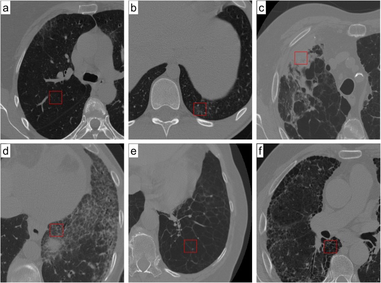
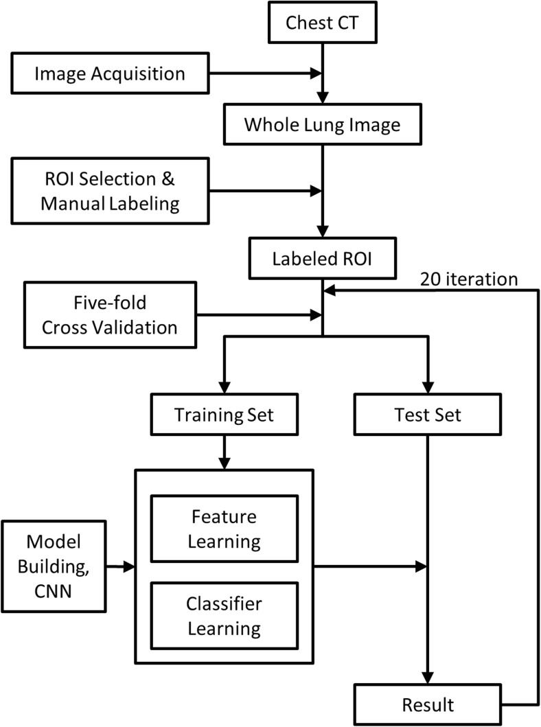
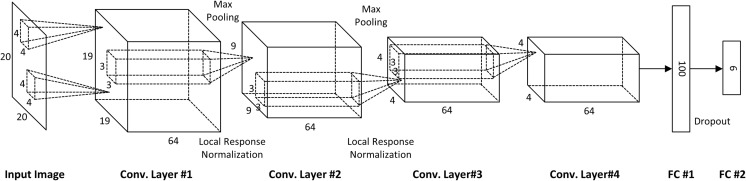
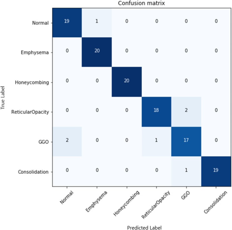
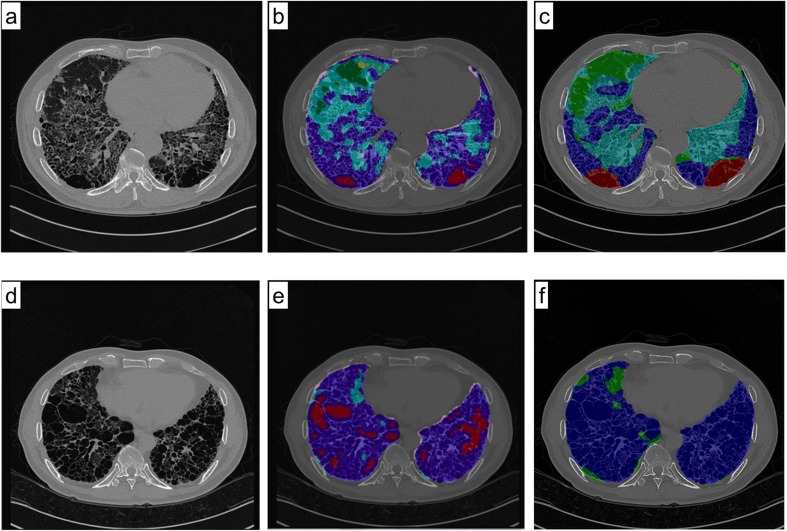
This study aimed to compare shallow and deep learning of classifying the patterns of interstitial lung diseases (ILDs). Using high-resolution computed tomography images, two experienced radiologists marked 1200 regions of interest (ROIs), in which 600 ROIs were each acquired using a GE or Siemens scanner and each group of 600 ROIs consisted of 100 ROIs for subregions that included normal and five regional pulmonary disease patterns (ground-glass opacity, consolidation, reticular opacity, emphysema, and honeycombing). We employed the convolution neural network (CNN) with six learnable layers that consisted of four convolution layers and two fully connected layers. The classification results were compared with the results classified by a shallow learning of a support vector machine (SVM). The CNN classifier showed significantly better performance for accuracy compared with that of the SVM classifier by 6-9%. As the convolution layer increases, the classification accuracy of the CNN showed better performance from 81.27 to 95.12%. Especially in the cases showing pathological ambiguity such as between normal and emphysema cases or between honeycombing and reticular opacity cases, the increment of the convolution layer greatly drops the misclassification rate between each case. Conclusively, the CNN classifier showed significantly greater accuracy than the SVM classifier, and the results implied structural characteristics that are inherent to the specific ILD patterns.
Keywords: Convolution neural network; Deep architecture; Interscanner variation; Interstitial lung disease; Support vector machine.
Figures


Similar articles
-
A support vector machine classifier reduces interscanner variation in the HRCT classification of regional disease pattern in diffuse lung disease: comparison to a Bayesian classifier.Med Phys. 2013 May;40(5):051912. doi: 10.1118/1.4802214. Med Phys. 2013. PMID: 23635282
-
Feasibility of automated quantification of regional disease patterns depicted on high-resolution computed tomography in patients with various diffuse lung diseases.Korean J Radiol. 2009 Sep-Oct;10(5):455-63. doi: 10.3348/kjr.2009.10.5.455. Epub 2009 Aug 25. Korean J Radiol. 2009. PMID: 19721830 Free PMC article.
-
Regional context-sensitive support vector machine classifier to improve automated identification of regional patterns of diffuse interstitial lung disease.J Digit Imaging. 2011 Dec;24(6):1133-40. doi: 10.1007/s10278-011-9367-0. J Digit Imaging. 2011. PMID: 21311944 Free PMC article.
-
Deep learning in interstitial lung disease-how long until daily practice.Eur Radiol. 2020 Nov;30(11):6285-6292. doi: 10.1007/s00330-020-06986-4. Epub 2020 Jun 14. Eur Radiol. 2020. PMID: 32537728 Free PMC article. Review.
-
Idiopathic interstitial pneumonias: CT features.Radiology. 2005 Jul;236(1):10-21. doi: 10.1148/radiol.2361031674. Radiology. 2005. PMID: 15987960 Review.
Cited by
-
Lung Segmentation on HRCT and Volumetric CT for Diffuse Interstitial Lung Disease Using Deep Convolutional Neural Networks.J Digit Imaging. 2019 Dec;32(6):1019-1026. doi: 10.1007/s10278-019-00254-8. J Digit Imaging. 2019. PMID: 31396776 Free PMC article.
-
Pattern classification of interstitial lung diseases from computed tomography images using a ResNet-based network with a split-transform-merge strategy and split attention.Phys Eng Sci Med. 2024 Jun;47(2):755-767. doi: 10.1007/s13246-024-01404-1. Epub 2024 Mar 4. Phys Eng Sci Med. 2024. PMID: 38436886
-
Recognition of Peripheral Lung Cancer and Focal Pneumonia on Chest Computed Tomography Images Based on Convolutional Neural Network.Technol Cancer Res Treat. 2022 Jan-Dec;21:15330338221085375. doi: 10.1177/15330338221085375. Technol Cancer Res Treat. 2022. PMID: 35293240 Free PMC article.
-
Generative Adversarial Network-Based Image Conversion Among Different Computed Tomography Protocols and Vendors: Effects on Accuracy and Variability in Quantifying Regional Disease Patterns of Interstitial Lung Disease.Korean J Radiol. 2023 Aug;24(8):807-820. doi: 10.3348/kjr.2023.0088. Korean J Radiol. 2023. PMID: 37500581 Free PMC article.
-
Prognostic value of deep learning-based fibrosis quantification on chest CT in idiopathic pulmonary fibrosis.Eur Radiol. 2023 May;33(5):3144-3155. doi: 10.1007/s00330-023-09534-y. Epub 2023 Mar 16. Eur Radiol. 2023. PMID: 36928568
References
Publication types
MeSH terms
LinkOut - more resources
Full Text Sources
Other Literature Sources
Medical
Research Materials