

doi: 10.1007/978-1-4939-7306-4_13.
Mapping DNA Breaks by Next-Generation Sequencing
Affiliations
- PMID: 29043624
- PMCID: PMC8057127
- DOI: 10.1007/978-1-4939-7306-4_13
Item in Clipboard
Mapping DNA Breaks by Next-Generation Sequencing
Methods Mol Biol.
2018.
Abstract
Here, we present two approaches to map DNA double-strand breaks (DSBs) and single-strand breaks (SSBs) in the genome of human cells. We named these methods respectively DSB-Seq and SSB-Seq. We tested the DSB and SSB-Seq in HCT1116, human colon cancer cells, and validated the results using the topoisomerase 2 (Top2)-poisoning agent etoposide (ETO). These methods are powerful tools for the direct detection of the physiological and pathological "breakome" of the DNA in human cells.
Keywords: DNA damage; Double-strand breaks (DSBs); Etoposide (ETO); Single-strand breaks (SSBs); Topoisomerase 2 (Top2).
Figures
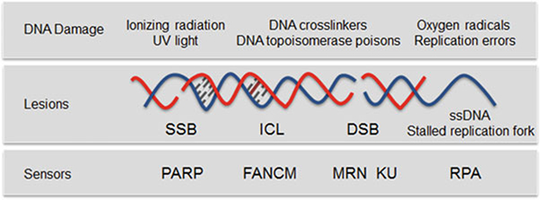
Types of DNA damage. Exogenous and endogenous DNA damaging agents generate various types of lesions including SSBs and DSBs. PARP predominantly acts as a sensor of SSB [17]. RPA binds to regions of single-stranded DNA (ssDNA) that are exposed to stalled replication forks or after DSB resection [18]. The multifunctional MRN complex and KU detect DSBs, FANCM is required for the DNA interstrand crosslink (ICL)-induced checkpoint response [19]. FANCM = Fanconi anemia complementation group M; ICL = interstrand crosslink; MRN = MRE11-RAD50-NBS1 complex; PARP = poly(ADP-ribose) polymerase; RPA = replication protein A
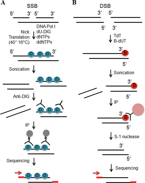
DNA breaks mapping workflow. (a) SSBs are labeled during nick translation using nucleotides covalently linked to digoxigenin (blue circle). The DNA is subsequently purified, sonicated and incubated with anti-digoxigenin antibody (anti-DIG). The immuno-precipitated DNA is sequenced. (b) 3′ tails of DSBs are ligated to biotinylated nucleotides (red circle). After sonication the labeled fragments are captured on streptavidin beads (pink circle). Tails are removed from released fragments and DNA is sequenced
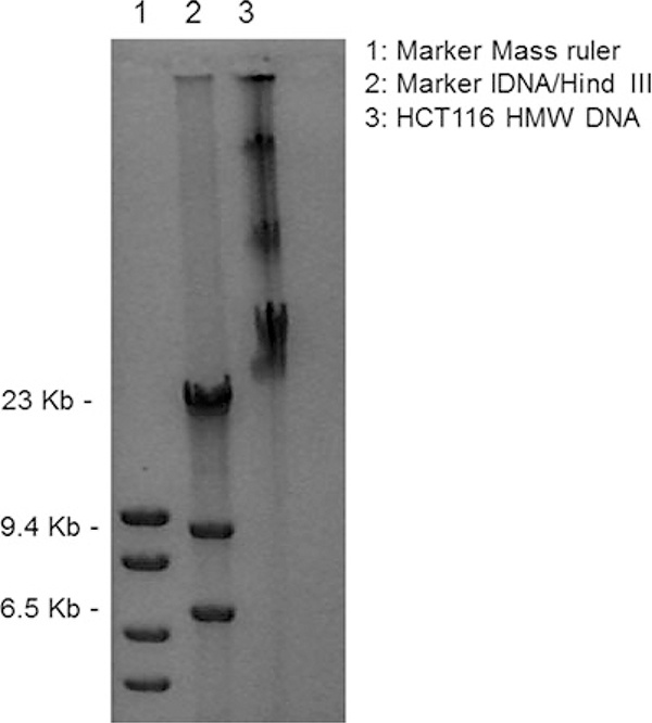
Representative example of High Molecular Weight (HMW) DNA after the purification steps described in Subheading 3.1. In lanes 1 and 2 we run two different markers. The numbers on the left refer to the molecular weight of the marker in lane 2
Similar articles
-
DNA break mapping reveals topoisomerase II activity genome-wide.Int J Mol Sci. 2014 Jul 23;15(7):13111-22. doi: 10.3390/ijms150713111. Int J Mol Sci. 2014. PMID: 25056547 Free PMC article.
-
A nucleotide resolution map of Top2-linked DNA breaks in the yeast and human genome.Nat Commun. 2019 Oct 24;10(1):4846. doi: 10.1038/s41467-019-12802-5. Nat Commun. 2019. PMID: 31649282 Free PMC article.
-
Bioflavonoids cause DNA double-strand breaks and chromosomal translocations through topoisomerase II-dependent and -independent mechanisms.Mutat Res Genet Toxicol Environ Mutagen. 2020 Jan;849:503144. doi: 10.1016/j.mrgentox.2020.503144. Epub 2020 Jan 22. Mutat Res Genet Toxicol Environ Mutagen. 2020. PMID: 32087851 Free PMC article.
-
Exploring the SSBreakome: genome-wide mapping of DNA single-strand breaks by next-generation sequencing.FEBS J. 2021 Jul;288(13):3948-3961. doi: 10.1111/febs.15568. Epub 2020 Oct 2. FEBS J. 2021. PMID: 32965079 Review.
-
Type II DNA Topoisomerases Cause Spontaneous Double-Strand Breaks in Genomic DNA.Genes (Basel). 2019 Oct 30;10(11):868. doi: 10.3390/genes10110868. Genes (Basel). 2019. PMID: 31671674 Free PMC article. Review.
Cited by
-
Pim kinase inhibitor co-treatment decreases alternative non-homologous end-joining DNA repair and genomic instability induced by topoisomerase 2 inhibitors in cells with FLT3 internal tandem duplication.Oncotarget. 2021 Aug 31;12(18):1763-1779. doi: 10.18632/oncotarget.28042. eCollection 2021 Aug 31. Oncotarget. 2021. PMID: 34504649 Free PMC article.
-
Endogenous DNA Double-Strand Breaks during DNA Transactions: Emerging Insights and Methods for Genome-Wide Profiling.Genes (Basel). 2018 Dec 14;9(12):632. doi: 10.3390/genes9120632. Genes (Basel). 2018. PMID: 30558210 Free PMC article. Review.
-
DNA damage and repair in differentiation of stem cells and cells of connective cell lineages: A trigger or a complication?Eur J Histochem. 2021 May 3;65(2):3236. doi: 10.4081/ejh.2021.3236. Eur J Histochem. 2021. PMID: 33942598 Free PMC article. Review.
-
Genome-wide detection of DNA double-strand breaks by in-suspension BLISS.Nat Protoc. 2020 Dec;15(12):3894-3941. doi: 10.1038/s41596-020-0397-2. Epub 2020 Nov 2. Nat Protoc. 2020. PMID: 33139954
References
-
- Blitzblau HG, Hochwagen A (2011) Genome-wide detection of meiotic DNA double-strand break hotspots using single-stranded DNA. Methods Mol Biol 745:47–63 - PubMed
MeSH terms
Substances
Grants and funding
LinkOut - more resources
Full Text Sources
Other Literature Sources
Miscellaneous
