

Determining residual adipose tissue characteristics with MRI in patients with various subtypes of lipodystrophy
- PMID: 29044029
- PMCID: PMC5669542
- DOI: 10.5152/dir.2017.17019
Determining residual adipose tissue characteristics with MRI in patients with various subtypes of lipodystrophy
Abstract
Purpose: We aimed to investigate residual adipose tissue with whole-body magnetic resonance imaging to differentiate between subtypes of lipodystrophy.
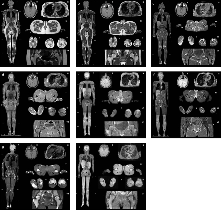
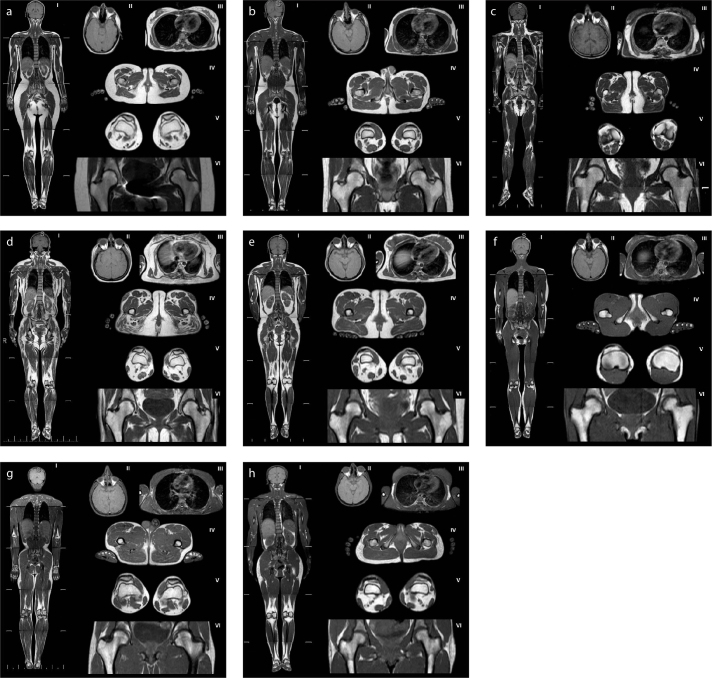
Methods: A total of 32 patients (12 with congenital generalized lipodystrophy [CGL], 1 with acquired generalized lipodystrophy [AGL], 12 with familial partial lipodystrophy [FPLD], and 7 with acquired partial lipodystrophy [APL]) were included.
Results: Despite generalized loss of metabolically active adipose tissue, patients with CGL1 caused by AGPAT2 mutations had a significant amount of residual adipose tissue in the scalp, earlobes, retro-orbital region, and palms and soles. No residual adipose tissue was noted particularly in the head and neck, palms and soles in CGL2 caused by BSCL2 mutations. CGL4 caused by mutations in the PTRF gene was characterized with well-preserved retro-orbital and bone marrow fat in the absence of any visible residual adipose tissue in other areas. No residual adipose tissue was observed in AGL. Despite loss of subcutaneous fat, periarticular adipose tissue was preserved in the lower limbs of patients with FPLD. Retro-orbital adipose tissue was surprisingly preserved in APL, although they lacked head and neck fat.
Conclusion: Lipodystrophies are a heterogeneous group of disorders characterized by generalized or partial loss of adipose tissue, which can be congenital or acquired. Our results suggest that residual adipose tissue characteristics can help distinguish different subtypes of lipodystrophy.
Conflict of interest statement
The authors declared no conflicts of interest.
Figures
References
-
- Garg A. Lipodystrophies. Am J Med. 2000;108:143–152. https://doi.org/10.1016/S0002-9343(99)00414-3. - DOI - PubMed
-
- Patni N, Garg A. Congenital generalized lipodystrophies-new insights into metabolic dysfunction. Nat Rev Endocrinol. 2015;11:522–534. https://doi.org/10.1038/nrendo.2015.123. - DOI - PMC - PubMed
-
- Agarwal AK, Arioglu E, De Almeida S, et al. AGPAT2 is mutated in congenital generalized lipodystrophy linked to chromosome 9q34. Nat Genet. 2002;31:21–23. https://doi.org/10.1038/ng880. - DOI - PubMed
-
- Magre J, Delepine M, Khallouf E, et al. Identification of the gene altered in Berardinelli-Seip congenital lipodystrophy on chromosome 11q13. Nat Genet. 2001;28:365–370. https://doi.org/10.1038/ng585. - DOI - PubMed
-
- Kim CA, Delepine M, Boutet E, et al. Association of a homozygous nonsense caveolin-1 mutation with Berardinelli-Seip congenital lipodystrophy. J Clin Endocrinol Metab. 2008;93:1129–1134. https://doi.org/10.1210/jc.2007-1328. - DOI - PubMed
MeSH terms
LinkOut - more resources
Full Text Sources
Other Literature Sources
Medical
