

Amalric sign: An augur of ophthalmic artery occlusion
- PMID: 29044080
- PMCID: PMC5678308
- DOI: 10.4103/ijo.IJO_42_17
Amalric sign: An augur of ophthalmic artery occlusion
Abstract
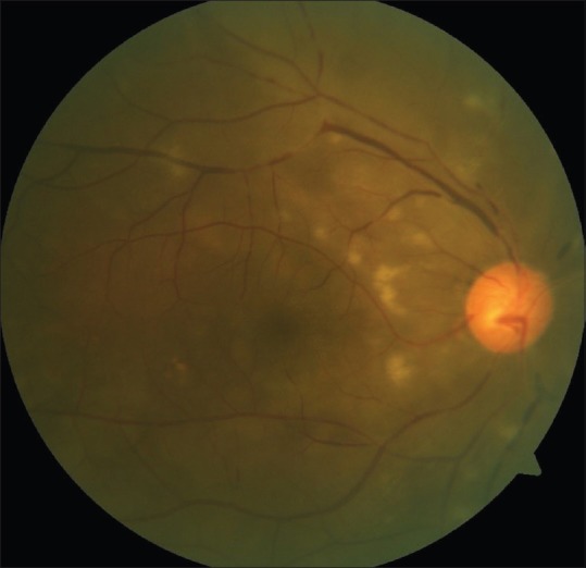
A 75-year-old man presented to us with sudden onset of profound vision loss in his right eye and was identified as suffering from an ophthalmic artery occlusion. Apart from the retinal whitening and box-carring of the retinal arteries, there were characteristic triangular patches of retinal whitening in the midperipheral temporal fundus indicating a previous lateral posterior choroidal artery occlusion. The patient was a chronic smoker and had dyslipidemia. The carotid Doppler study showed complete occlusion of the internal carotid artery. The presence of these triangular patches of retinal whitening or amalric sign can therefore herald a more proximal vessel occlusion. Hence such patients require evaluation on an emergency basis. The characteristic features of the patches on fluorescein angiography and indocyanine green angiography are discussed here.
Conflict of interest statement
There are no conflicts of interest.
Figures


Similar articles
-
[Spontaneous visual recovery following a central retinal artery occlusion in a patient with a cilioretinal artery].Orv Hetil. 2019 Jul;160(29):1146-1152. doi: 10.1556/650.2019.31434. Orv Hetil. 2019. PMID: 31303015 Hungarian.
-
CENTRAL RETINAL ARTERY OCCLUSION WITH DOUBLE CILIORETINAL ARTERY SPARING.Retin Cases Brief Rep. 2019 Winter;13(1):75-78. doi: 10.1097/ICB.0000000000000537. Retin Cases Brief Rep. 2019. PMID: 28085758
-
Evaluation of bilateral central retinal artery occlusions with optical coherence tomography-based microangiography: a case report.J Med Case Rep. 2016 Nov 1;10(1):307. doi: 10.1186/s13256-016-1095-0. J Med Case Rep. 2016. PMID: 27802835 Free PMC article.
-
STRANGULATION-INDUCED CENTRAL RETINAL ARTERY OCCLUSION: CASE REPORT AND REVIEW OF THE LITERATURE.Retin Cases Brief Rep. 2017 Summer;11(3):258-260. doi: 10.1097/ICB.0000000000000334. Retin Cases Brief Rep. 2017. PMID: 27337704 Review.
-
Bilateral Combined Central Retinal Artery and Vein Occlusion in a Child with Purtscher Retinopathy: A Case Report and Review of the Literature.Middle East Afr J Ophthalmol. 2024 Dec 2;30(4):274-280. doi: 10.4103/meajo.meajo_170_23. eCollection 2023 Oct-Dec. Middle East Afr J Ophthalmol. 2024. PMID: 39959587 Free PMC article. Review.
Cited by
-
Novel ultrawide field fundus fluorescein angiographic findings in a patient of Takayasu arteritis on immunosuppression.Indian J Ophthalmol. 2020 Jan;68(1):238-240. doi: 10.4103/ijo.IJO_455_19. Indian J Ophthalmol. 2020. PMID: 31856538 Free PMC article.
-
Amalric choroidal infarction, retinal artery occlusion, and ischemic optic neuropathy: Delayed presentations of traumatic internal carotid artery dissection.Am J Ophthalmol Case Rep. 2024 Oct 11;36:102193. doi: 10.1016/j.ajoc.2024.102193. eCollection 2024 Dec. Am J Ophthalmol Case Rep. 2024. PMID: 39498145 Free PMC article.
References
-
- Amalric P. Acute choroidal ischaemia. Trans Ophthalmol Soc U K. 1971;91:305–22. - PubMed
-
- Reddy S, Goldman DR, Hubschman JP, Kaines A, Sarraf D. Cocaine and choroidal infarction, revisiting the triangular sign of amalric. Retin Cases Brief Rep. 2011;5:91–3. - PubMed
-
- Hsu CT, Kerrison JB, Miller NR, Goldberg MF. Choroidal infarction, anterior ischemic optic neuropathy, and central retinal artery occlusion from polyarteritis nodosa. Retina. 2001;21:348–51. - PubMed
-
- Hayreh SS. Posterior ciliary artery circulation in health and disease: The Weisenfeld lecture. Invest Ophthalmol Vis Sci. 2004;45:749–57. 748. - PubMed
Publication types
MeSH terms
LinkOut - more resources
Full Text Sources
Other Literature Sources
