

Case Reports
doi: 10.4103/ijo.IJO_345_17.
Management of idiopathic intracranial hypertension in an infant with bilateral congenital cataract and associated comitant sensory esotropia
Affiliations
- PMID: 29044088
- PMCID: PMC5678316
- DOI: 10.4103/ijo.IJO_345_17
Item in Clipboard
Case Reports
Management of idiopathic intracranial hypertension in an infant with bilateral congenital cataract and associated comitant sensory esotropia
Indian J Ophthalmol.
2017 Oct.
Abstract
In this report, we describe the management of a child with bilateral cataract, nystagmus, and comitant sensory esotropia. Routine ultrasonography done before cataract surgery revealed bilateral disc edema confirmed as idiopathic intracranial hypertension by a pediatric neurologist. The primary intervention for cataract surgery was followed by nonresolving papilledema, despite maximum medical therapy. To salvage the optic nerve function in a nonverbal child, bilateral optic nerve sheath decompression was planned with simultaneous medial rectus recessions for the persistent esotropia with the satisfactory postoperative outcome.
Conflict of interest statement
There are no conflicts of interest.
Figures
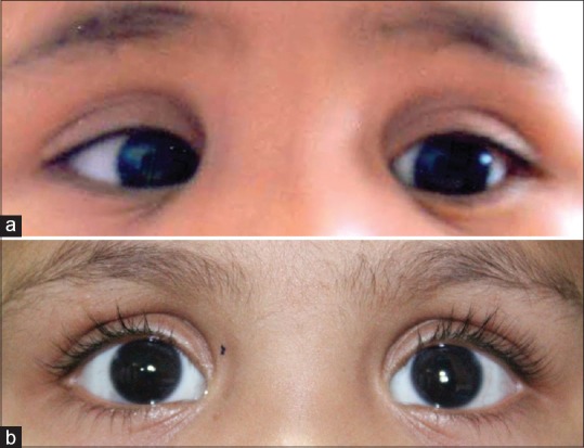
Clinical photograph of child; (a) preoperatively showing right esotropia with psuedophakia in both eyes, (b) postoperatively, photograph showing orthotropia and pseudophakia in both eyes

B-scan right (left image) and left eye (right image); (a) at presentation showing optic nerve head (thick arrow) with size of 5 mm in right eye and 5.30 mm in left eye and an echolucent crescent due to the sub-arachnoid fluid around the optic nerve (thin arrow), intraoperative image showing, (b) distended optic nerve sheath (arrowhead) and short posterior ciliary vessels (thin arrow) over optic nerve, (c) B-scan right (left image) and left eye (right image); postoptic nerve sheath decompression showing decrease in optic nerve head size (thick arrow) with resolution of sub-arachnoid fluid around optic nerve

Magnetic resonance imaging of brain (T2 sagittal scan) showing distended optic nerve sheaths indicated by white arrows in both eyes
Similar articles
-
A case of pediatric idiopathic intracranial hypertension presenting with divergence insufficiency.Korean J Ophthalmol. 2011 Aug;25(4):289-93. doi: 10.3341/kjo.2011.25.4.289. Epub 2011 Jul 22. Korean J Ophthalmol. 2011. PMID: 21860580 Free PMC article.
-
Results of bilateral medial rectus recession for comitant esotropia in patients with developmental delay.Strabismus. 2014 Sep;22(3):138-42. doi: 10.3109/09273972.2014.907814. Epub 2014 May 5. Strabismus. 2014. PMID: 24798741
-
Findings of magnetic resonance imaging after optic nerve sheath decompression in patients with idiopathic intracranial hypertension.Am J Ophthalmol. 2007 Sep;144(3):429-435. doi: 10.1016/j.ajo.2007.05.034. Epub 2007 Jul 19. Am J Ophthalmol. 2007. PMID: 17640608
-
Divergence Insufficiency Esotropia: Surgical Treatment.Am Orthopt J. 2015;65:35-9. doi: 10.3368/aoj.65.1.35. Am Orthopt J. 2015. PMID: 26564924 Free PMC article. Review.
-
Botulinum toxin treatment for esotropia.Am Orthopt J. 2013;63:29-31. doi: 10.3368/aoj.63.1.29. Am Orthopt J. 2013. PMID: 24260805 Review.
Cited by
-
Idiopathic Intracranial Hypertension in Neonates, Infants, and Toddlers.J Clin Med. 2025 Jul 17;14(14):5084. doi: 10.3390/jcm14145084. J Clin Med. 2025. PMID: 40725776 Free PMC article. Review.
-
Inaccuracy of idiopathic intracranial hypertension diagnosis in case reports.Eye (Lond). 2023 Oct;37(15):3243-3248. doi: 10.1038/s41433-023-02499-8. Epub 2023 Mar 16. Eye (Lond). 2023. PMID: 36928224 Free PMC article. Review.
-
Infantile idiopathic intracranial hypertension: case report and review of the literature.Ital J Pediatr. 2022 Jan 10;48(1):3. doi: 10.1186/s13052-021-01191-5. Ital J Pediatr. 2022. PMID: 35012609 Free PMC article. Review.
References
-
- Matalia J, Shirke S, Kekatpure M. An alternate technique for assessing optic nerve in papilledema by ultrasound B scan. Am J Emerg Med. 2015;33:971–3. - PubMed
-
- Stone MB. Ultrasound diagnosis of papilledema and increased intracranial pressure in pseudotumor cerebri. Am J Emerg Med. 2009;27:376.e1–376.e2. - PubMed
-
- Wall M. Idiopathic intracranial hypertension. Neurol Clin. 1991;9:73–95. - PubMed
-
- Ko MW, Liu GT. Pediatric idiopathic intracranial hypertension (pseudotumor cerebri) Horm Res Paediatr. 2010;74:381–9. - PubMed
-
- Phillips PH. Pediatric pseudotumor cerebri. Int Ophthalmol Clin. 2012;52:51–9, xii. - PubMed
Publication types
MeSH terms
LinkOut - more resources
Full Text Sources
Other Literature Sources
Medical
