. 2017 Oct 18;8(1):1028.
doi: 10.1038/s41467-017-01022-4.

DNA-binding of the Tet-transactivator curtails antigen-induced lymphocyte activation in mice

Affiliations

DNA-binding of the Tet-transactivator curtails antigen-induced lymphocyte activation in mice

Eleonora Ottina et al. Nat Commun. .

Abstract

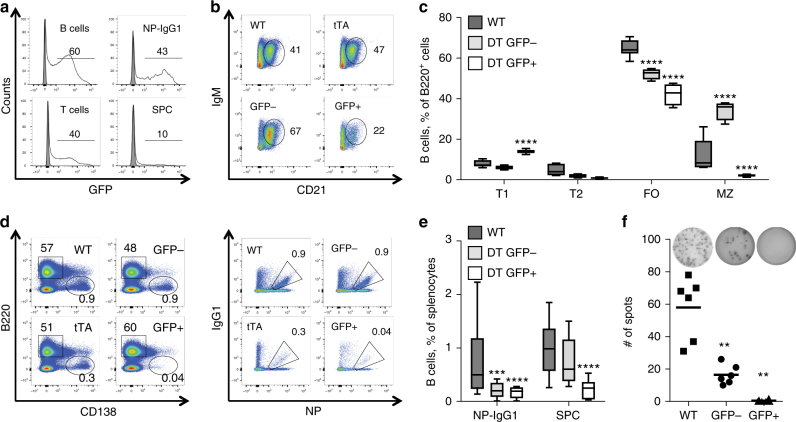
The Tet-On/Off system for conditional transgene expression constitutes state-of-the-art technology to study gene function by facilitating inducible expression in a timed and reversible manner. Several studies documented the suitability and versatility of this system to trace lymphocyte fate and to conditionally express oncogenes or silence tumour suppressor genes in vivo. Here, we show that expression of the tetracycline/doxycycline-controlled Tet-transactivator, while tolerated well during development and in immunologically unchallenged animals, impairs the expansion of antigen-stimulated T and B cells and thereby curtails adaptive immune responses in vivo. Transactivator-mediated cytotoxicity depends on DNA binding, but can be overcome by BCL2 overexpression, suggesting that apoptosis induction upon lymphocyte activation limits cellular and humoral immune responses. Our findings suggest a possible system-intrinsic biological bias of the Tet-On/Off system in vivo that will favour the outgrowth of apoptosis resistant clones, thus possibly confounding data published using such systems.


Conflict of interest statement

The authors declare no competing financial interests.

Figures

Fig. 1
Fig. 1
tTA transactivator expression compromises germinal centre formation and development of antigen-specific antibody-secreting cells. Flow cytometric analysis of splenocytes from vav-tTA mice or littermate controls at day 7 post immunization with NP-KLH. a Percentages of transitional type 1 (T1) B cells (B220high IgMhigh CD23low CD21low), transitional type 2 (T2) B cells (B220high IgMhigh CD23+ CD21high), follicular (FO) B cells (B220high IgMlow CD23+ CD21low) and marginal zone (MZ) B cells (B220high IgMhigh CD23low CD21high). For the gating strategy, please see the Supplementary Fig. 9a. Data are presented as box-and-whiskers diagram with median and interquartile range (vav-tTA n = 10; littermate controls n = 7). **P ≤ 0.01, vav-tTA vs. wild-type (WT), one-way ANOVA with Bonferroni correction for multiple comparisons. b Representative staining with B220- and CD138-specific antibodies to identify plasma cells (left) or NP-specific IgG1+ germinal centre B cells (left), defined as IgM IgD Gr1 CD138 B220+ IgG1-NP+. For the gating strategy, please see the Supplementary Fig. 9b. c Frequencies of splenic plasma cells (SPC) (B220low CD138+) and germinal centre NP-specific isotype-switched B cells (IgM IgD Gr1 CD138 B220+ NP+ IgG1+). Data are presented as box-and-whiskers diagram with median and interquartile range (vav-tTA n = 14; littermate controls n = 13). **P ≤ 0.01, vav-tTA vs. wild-type (WT), one-way ANOVA with Bonferroni correction for multiple comparisons. d Frequencies of total NP-specific IgG1-secreting cells in spleen determined by ELISPOT assay. Data are presented as scatter plots with mean; each dot represents data from a single immunized mouse as a mean of assays in two replicate wells, summarizing three independent experiments (n = 4–6 per genotype and immunization). ****P < 0.0001, vav-tTA vs. WT, two-sample Kolmogorov–Smirnov test
Fig. 2
Fig. 2
tTA expression level correlates with the severity of impairment of B cell activation. Flow cytometric analysis of splenocytes from TRE_Ren/vav-tTA or TRE-FF/vav-tTA double transgenic (DT) mice or littermate controls at day 7 after immunization with NP-KLH. a GFP reporter expression in B cells (B220+), T cells (TCRβ+), plasma cells (B220low CD138+) and germinal centre NP-specific B cells (IgM IgD Gr1 CD138 B220+ NP+ IgG1+) splenocytes of DT mice, open histograms, and wt mice, filled histograms; percentages given refer to GFP+ cells. b Representative dot plots of wt, vav-tTA and DT splenocytes gated on B220high and CD23low and c quantification of transitional type 1 (T1) B cells (B220high IgMhigh CD23low CD21low), transitional type 2 (T2) B cells (B220high IgMhigh CD23+ CD21high), follicular (FO) B cells (B220high IgMlow CD23+ CD21low) and marginal zone (MZ) B cells (B220high IgMhigh CD23low CD21high) of wt and DT mice sorted on the basis of the GFP expression level. For the gating strategy, please see the Supplementary Fig. 9a. Data are presented as box-and-whiskers diagram with median and interquartile range (DT mice n = 7; littermate controls n = 7). ***P ≤ 0.001, ****P ≤ 0.0001, GFP+ or GFP vs. controls, two-way ANOVA with Bonferroni correction for multiple comparisons. d Representative plasma cell staining, using B220 and CD138 specific antibodies (left) and NP-specific IgM IgD Gr1 CD138 B220+ IgG1-NP+ (right) germinal centre B cells, quantified in e. For the gating strategy, please see the Supplementary Fig. 9b. Data are presented as box-and-whiskers diagram with median and interquartile range (DT mice n = 9; littermate controls n = 9). **P ≤ 0.01,***P ≤ 0.001, ****P ≤ 0.0001, GFP+ and GFP vs. controls, two-way ANOVA with Bonferroni correction for multiple comparisons. f Frequencies of total NP-specific IgG1-secreting cells among FACS-sorted splenocytes as determined by ELISPOT assay. Each dot represents data from a single mouse as mean of assays in replicate wells from two independent experiments using three mice per genotype. Data are presented as scatter plots with mean. **P < 0.01, GFP+ vs. GFP subsets, two-sample Kolmogorov–Smirnov test
Fig. 3
Fig. 3
tTA transactivator expression impairs development of T cell immunity during acute viral infection. Flow cytometric analysis of splenocytes from vav-tTA mice (n = 4) or littermate controls (n = 4) at day 8 after infection with LCMV strain WE. a Percentages of CD4+ and CD8+ T cells in spleens of infected mice. b Flow cytometric analysis of LCMV-specific CD8+ T cells in spleens of wt and vav-tTA mice upon infection. Cells were stained with antibodies against CD8, PD-1 and tetramers for low-affinity and high-affinity peptides of LCMV proteins: H-2Db/GP33 (KAVYNFATM), H-2Db/GP276 (SGVENPGGYCL) and H-2Db/NP396 (FQPQNGQFI), conjugated with streptavidin APC (left); IFNγ and TNF production in virus-specific CD8+ T cells after a 4 h in vitro restimulation of splenocytes with the cognate peptide was assessed by intracellular staining and flow cytometric analysis (right). For the gating strategy, please see the Supplementary Fig. 9c, d. c Quantification of LCMV-reactive tetramer-positive CD8+ T cells. d Quantification of cytokine-positive tetramer positive CD8+ T cells after in vitro restimulation. Data are presented as box-and-whiskers diagram with median and interquartile range of four mice per genotype. **P < 0.01, ***P ≤ 0.001, ****P ≤ 0.0001, two-way ANOVA with Bonferroni correction for multiple comparisons
Fig. 4
Fig. 4
The tTA transactivator induced loss of germinal centre and splenic plasma cells in immunized mice is B cell-autonomous. Flow cytometric analysis of splenocytes from Eμ-tTA mice (n = 6) or littermate control (n = 6) at day 7 after immunization with NP-KLH. a Representative stainings with antibodies against B220 and CD138 markers to identify plasma cells (left) or NP-specific IgG1+ germinal centre B cells (left) defined as IgM IgD Gr1 CD138 B220+ IgG1-NP+. For the gating strategy, please see the Supplementary Fig. 9b. b Frequencies of germinal centre NP-specific isotype-switched B cells (IgM IgD Gr1 CD138 B220+ IgG1-NP+) and splenic plasma cells (B220low CD138+). Data are presented as box-and-whiskers diagram with median and interquartile range. **P ≤ 0.01, Eμ-tTA vs. WT, two-way ANOVA with Bonferroni correction for multiple comparisons. c Frequencies of total NP-specific IgG1-secreting cells in spleen as determined by ELISPOT assay. Each dot represents data from a single mouse as the mean of assays in replicate wells, with four mice in each group, and summarizes two independent experiments. Data are presented as scatter plots with mean. ***P < 0.001, Eμ-tTA vs. WT, two-sample Kolmogorov–Smirnov test
Fig. 5
Fig. 5
Tet transactivator-mediated cytotoxicity in antigen stimulated B cells depends on DNA binding. a Representative staining (left) and frequencies (right) of flow cytometric analysis of splenic plasma cells (B220low CD138+) from vav-tTA (n = 10), CAG-rtTA (n = 8) or littermate control mice (n = 9) at day 7 after immunization with NP-KLH or vav-tTA mice (n = 5), CAG-rtTA mice (n = 9) or littermate control (n = 8) fed with doxycycline-containing food for 7 days prior and during the 7 days of NP-KLH challenge. b Representative FACS analysis (left) and frequencies (right) of germinal centre NP-specific isotype-switched B cells (IgM IgD Gr1 CD138 B220+ IgG1-NP+) from the mice of the indicated genotype as in a. For the gating strategy, please see the Supplementary Fig. 9b. Data are presented as box-and-whiskers diagram with median and interquartile range. *P ≤ 0.05, **P ≤ 0.01, ***P < 0.001, one-way ANOVA
Fig. 6
Fig. 6
Tet transactivator-mediated cytotoxicity in antibody-secreting plasma cells depends on DNA binding. a Frequencies of total NP-specific IgG1-secreting cells in spleen as determined by ELISPOT assays on splenocytes from mice of the indicated genotypes at day 7 after immunization with NP-KLH or fed with doxycycline-containing food for 7 days prior and during the 7 days of NP-KLH challenge. Each dot represents data from a single mouse as mean of results from assays in replicate wells, with three to four mice in each group, and summarizes three independent experiments. Data are presented as scatter plots with mean. ****P < 0.0001, vs. WT or WT on DOX food, two-sample Kolmogorov–Smirnov test. b Representative H&E-stained splenic sections from the above-mentioned mice, scale bars represent 1 mm or 200 μm in the magnified views. White line demarcates the area used by the random forest classifier of Ilastik (see Methods section) for (c) quantification of the cumulative follicular area over total spleen section area. Data are presented as scatter plots with mean. **P < 0.01 vav-tTA vs. wild type and ****P < 0.0001, vav-tTA vs. vav-tTA on DOX-containing food, two-sample Kolmogorov–Smirnov test
Fig. 7
Fig. 7
Overexpression of Bcl-2 prevents the transactivator-mediated cytotoxicity in antigen stimulated B cells. Flow cytometric analysis of splenocytes from vav-Bcl-2/vav-tTA double transgenic mice (n = 10) or vav-Bcl-2 littermate controls (n = 10) at day 7 after immunization with NP-KLH. a Representative staining using B220 and CD138-secific antibodies to identify plasma cells (left) and NP-specific IgG1+ germinal centre B cells as IgM IgD Gr1 CD138 B220+ IgG1-NP (right). For the gating strategy, please see the Supplementary Fig. 9b. b Frequency of GC and splenic plasma cells. Data are presented as box-and-whiskers diagram with median and interquartile range. c Frequencies of total NP-specific IgG1-secreting cells in spleens as determined by ELISPOT assays. Each dot represents data from a single mouse as mean of results from assays in replicate wells and five mice in each group, summarizing two independent experiments. Data are presented as scatter plot. d H&E-stained splenic sections from the above-mentioned mice, white line represent the follicle area used by the random forest classifier of Ilastik and e quantification of the cumulative follicular area over total spleen section area. Scale bars represent 1 mm or 200 μm in the magnified views. No significant differences were observed

Similar articles

Cited by

References

    1. Bouabe H, Okkenhaug K. Gene targeting in mice: a review. Method. Mol. Biol. 2013;1064:315–336. doi: 10.1007/978-1-62703-601-6_23. - DOI - PMC - PubMed
    1. Capecchi MR. Gene targeting in mice: functional analysis of the mammalian genome for the twenty-first century. Nat. Rev. Genet. 2005;6:507–512. doi: 10.1038/nrg1619. - DOI - PubMed
    1. Lindsten T, et al. The combined functions of proapoptotic Bcl-2 family members Bak and Bax are essential for normal development of multiple tissues. Mol. Cell. 2000;6:1389–1399. doi: 10.1016/S1097-2765(00)00136-2. - DOI - PMC - PubMed
    1. Labi V, et al. Deregulated cell death and lymphocyte homeostasis cause premature lethality in mice lacking the BH3-only proteins Bim and Bmf. Blood. 2014;123:2652–2662. doi: 10.1182/blood-2013-11-537217. - DOI - PMC - PubMed
    1. Rossi A, et al. Genetic compensation induced by deleterious mutations but not gene knockdowns. Nature. 2015;524:230–233. doi: 10.1038/nature14580. - DOI - PubMed

Publication types

LinkOut - more resources