. 2017 Oct 18;7(1):13480.
doi: 10.1038/s41598-017-13620-9.

Modeling and Preventing Progressive Hearing Loss in Usher Syndrome III

Affiliations

Modeling and Preventing Progressive Hearing Loss in Usher Syndrome III

Ruishuang Geng et al. Sci Rep. .

Abstract

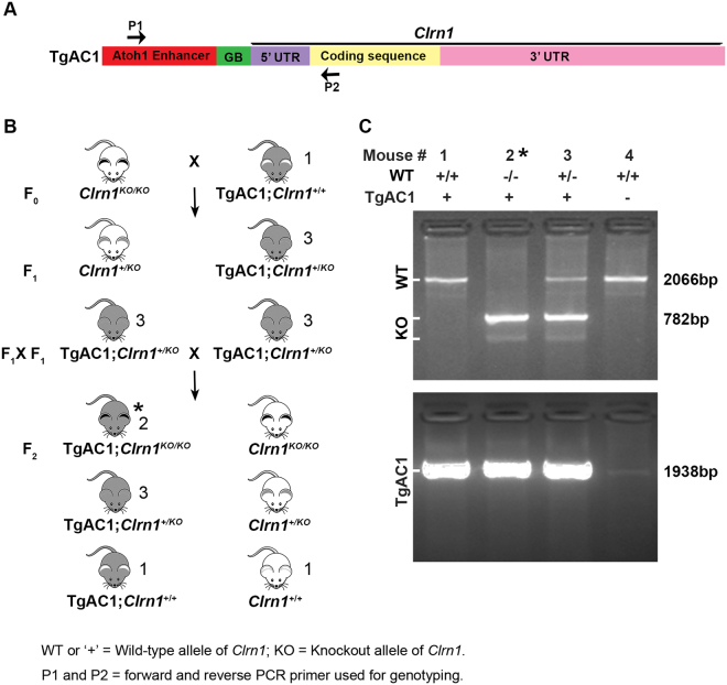
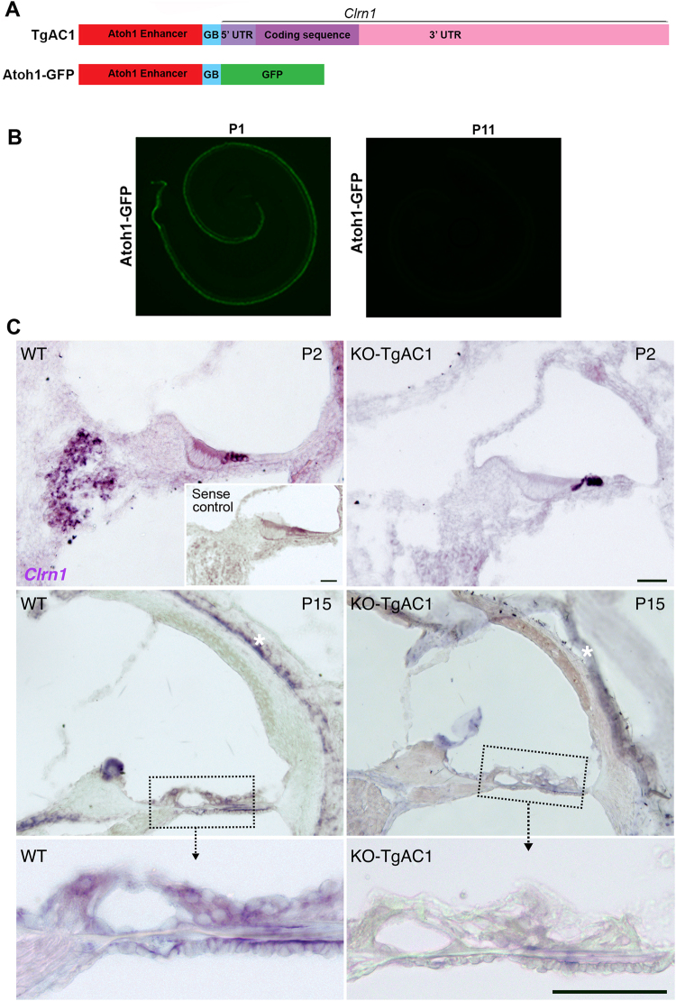
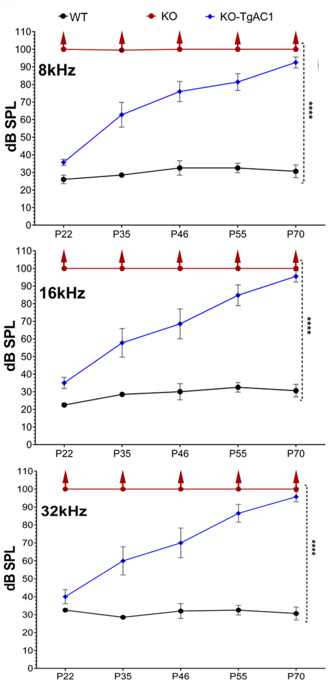
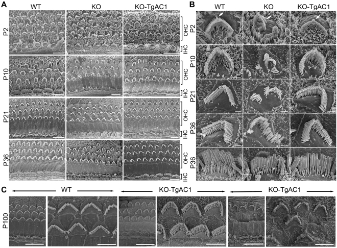
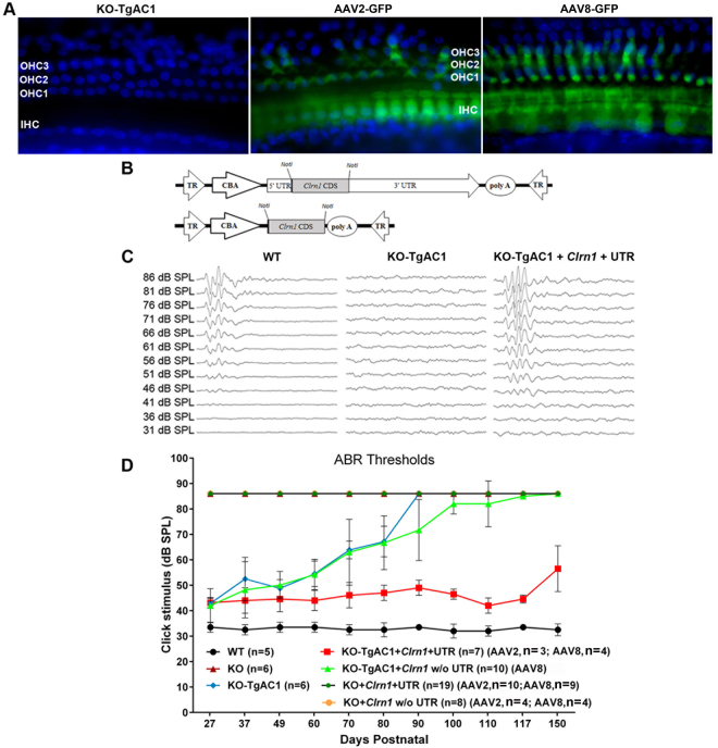
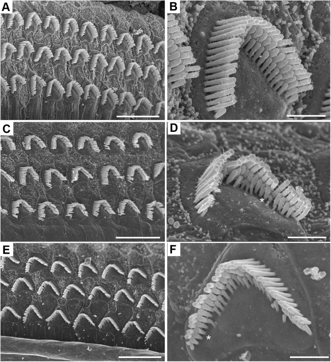
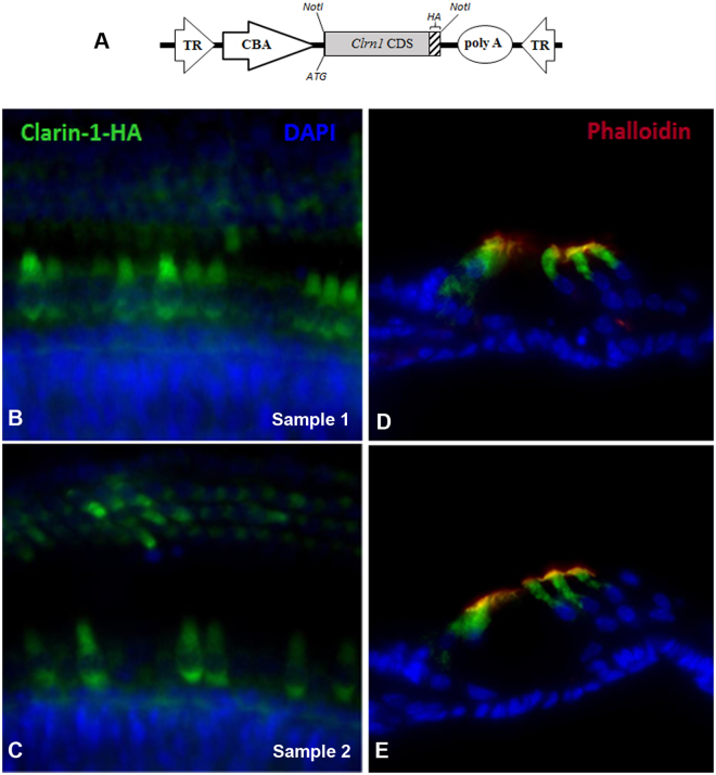
Usher syndrome type III (USH3) characterized by progressive loss of vision and hearing is caused by mutations in the clarin-1 gene (CLRN1). Clrn1 knockout (KO) mice develop hair cell defects by postnatal day 2 (P2) and are deaf by P21-P25. Early onset profound hearing loss in KO mice and lack of information about the cochlear cell type that requires Clrn1 expression pose challenges to therapeutic investigation. We generated KO mice harboring a transgene, TgAC1, consisting of Clrn1-UTR (Clrn1 cDNA including its 5' and 3' UTR) under the control of regulatory elements (Atoh1 3' enhancer/β-globin basal promoter) to direct expression of Clrn1 in hair cells during development and down regulate it postnatally. The KO-TgAC1 mice displayed delayed onset progressive hearing loss associated with deterioration of the hair bundle structure, leading to the hypothesis that hair cell expression of Clrn1 is essential for postnatal preservation of hair cell structure and hearing. Consistent with that hypothesis, perinatal transfection of hair cells in KO-TgAC1 mice with a single injection of AAV-Clrn1-UTR vector showed correlative preservation of the hair bundle structure and hearing through adult life. Further, the efficacy of AAV-Clrn1 vector was significantly attenuated, revealing the potential importance of UTR in gene therapy.


Conflict of interest statement

The authors declare the following competing financial interests: The mouse Clrn1 cDNA used to generate the transgenic mouse TgAC1 and the human ortholog are protected by a provisional US patent # 62/076,114 and 62/158,846 to K.N.A. WWH and the University of Florida have a financial interest in the use of AAV therapies and own equity in a company (AGTC Inc.) that might, in the future, commercialize some aspects of this work.

Figures

Figure 1
Figure 1
Generation of KO-TgAC1 mice. (A) A schematic of a transgene construct. The transgene construct is composed of an Atoh1 enhancer sequence fused to the beta globin basal (GB) promoter sequence. The Clrn1 cDNA fused downstream of the Atoh1 regulatory elements is composed of the 5′ UTR sequence, the coding sequence (isoform 2) and the 3′ UTR sequence. (B) The breeding scheme used to generate KO-TgAC1 mice. The expanded symbol for the KO-TgAC1 mice is “TgAC1; Clrn1 KO/KO”, marked by an asterisk in the F2 generation. The number next to the mouse corresponds to the lane number in panel C. (C) PCR-based genotyping to identify the WT (2066 bp), KO (782 bp) and TgAC1 (1938 bp) allele of Clrn1. *F2 mice with the desired genotype, TgAC1; Clrn1 KO/KO.
Figure 2
Figure 2
Mouse clarin-1 transcript. 5′ UTR, 172 bp (black font); coding sequence, 696 bp (underlined) start ‘ATG’ and stop ‘TGA’ codon indicated (bold, red font); exon 1 (red font), exon 3 (green font) and exon 4 (blue font); 3′UTR, 2158 bp (black font, italicized text; lower case italicized text indicates additional bases found in the GenBank sequence (Accession number: NM_153384.3) not included in the TgAC1 construct.
Figure 3
Figure 3
Conditional expression of Clrn1 in KO-TgAC1 mice. (A) The schematic diagram shows Atoh1-Clrn1 and Atoh1-GFP transgene constructs. (B) GFP expression in the organ of Corti at P1 and P11 from the Atoh1-GFP transgenic line. (C) Clrn1 ISH on cochlear duct sections from WT or KO-TgAC1 mice at P2 (top row) and P15 (second and bottom row). The inset (sense probe, top row) and asterisks (labyrinth cartilage and tectorial membrane, second row) show background staining. Scale bar = 50 µM.
Figure 4
Figure 4
Progressive hearing loss in Clrn1 KO mice harboring the TgAC1 transgene. To monitor hearing over time, ABR thresholds were recorded longitudinally from the same set of mice in each group (genotype). ABR thresholds of WT (n = 5), KO (n = 10) and KO-TgAC1 (n = 10) mice from P22 to P70 at 8, 16 and 32 kHz. ****p < 0.0001. No ABR was generated in the KO mice at the highest intensity (100 dB SPL) tested; the upward arrow indicates that the threshold is greater than 100 db SPL. The error bar in each panel represents standard error of the mean.
Figure 5
Figure 5
Delayed onset of hair cell damage in KO-TgAC1. (A) FESEM images of WT, KO and KO-TgAC1 organ of Corti from mid-cochlear regions. At all ages illustrated, the hair bundle morphology appears disrupted in all rows of the OHCs in the KO, whereas the hair bundle morphology in the KO-TgAC1 mice is grossly comparable to the WT specimen. Scale bars = 10 μm. (B) At higher magnification, disturbances of the hair bundles apparent in the KO specimens are much reduced in the KO-TgAC1 specimens. The upper four rows show OHCs at P2-P36. The KO shows disturbed bundles with splits in the rows and missing stereocilia, and distorted apical surfaces, while the KO-TgAC1 has a near normal appearance. Some of the short stereocilia in all three mice are missing (*), an effect which is greater in KO-TgAC1 and greater still in the KO compared with the WT. The bottom row shows IHCs at P36. In both the KO and KO-TgAC1 mice, short stereocilia are missing compared with the WT, but overall the hair bundle in KO-TgAC1 mice is more similar to the WT than the KO. Scale bars = 1 μm. (C) At P100, the majority of the OHCs along the cochlear turn are severely affected (panels to the right) with a few patches of the organ of Corti that appear to be less severely affected (panels to the center) compared to the WT (panels to the left). The organ of Corti from the mid-basal turn of the cochlea is shown here. All mice are maintained in the C57BL/6J background; five mice (n = 5) per each genotype were used for the SEM analysis. Scale bars = 5 μm.
Figure 6
Figure 6
AAV-mediated gene therapy in the KO-TgAC1 USH3 model. (A) Surface preparation of the organ of Corti from KO-TgAC1 mice stained with DAPI (left panel), transfected with AAV2 -GFP (middle panel) or AAV8-GFP (right panel). AAV2-GFP and AAV8-GFP were used to assess viral delivery to the cochlea of KO-TgAC1. High titer stock (>1013 vg/ml) of AAV2 or 8-GFP was injected through the RWM of the KO-TgAC1 mice at P1-P3, and the organ of Corti was examined for GFP expression in the cochlea whole mount at P10 by immunofluorescence using anti-GFP antibody. Almost all IHCs were GFP positive (green), whereas a mosaic pattern of GFP positive cells was observed in the three rows of OHCs. Representative specimens from the mid-basal turn of the cochlea from mice injected with AAV2-GFP or AAV8-GFP  are shown in the middle and right panels. (BD) Hearing restoration in the KO-TgAC1 mice. (B) Schematic representation of AAV vector constructs used in gene rescue experiments. The AAV expression cassette contains the mouse clarin-1 coding sequence (CDS) with or without its UTR sequences. Both constructs were packaged in either serotype 2 or serotype 8 AAV capsids. The Clrn1 cDNA expression in each vector is driven by the ubiquitous small CBA promoter. The expression cassette is flanked by inverted terminal repeats (TR) of AAV serotype 2; poly-A, SV40 polyadenylation signal. (C) Representative ABR traces in response to click stimulus from WT, KO-TgAC1 and rescued KO-TgAC1 mice following delivery of 2 μl AAV2 or AAV8-Clrn1-UTR construct. At P100, waveforms from WT and KO-TgAC1 mice injected with AAV2-Clrn1-UTR appear normal and similar, while non-injected KO-TgAC1 mice show no ABR responses at the sound levels tested. (D) AAV2 or AAV8 containing the Clrn1 gene, with or without the UTR sequence, were directly injected through the RWM of the KO or KO-TgAC1 mice at P1-P3 when the hair cells in KO-TgAC1 mice are comparable to the WT hair cells. To monitor preservation or loss of hearing over time, ABR thresholds were recorded from the same set of mice in each group (genotype). ABRs evoked with click stimulus recorded longitudinally from the same mice 4 to 22 weeks after birth (P27 to P150). The KO-TgAC1 mice injected with AAV2 or AAV8-Clrn1-UTR (n = 7) (red line) showed significant preservation of hearing through their adult life compared to the KO-TgAC1 mice injected with Clrn1 without UTR (n = 10) or non-injected sham surgery KO-TgAC1 (n = 6). There was no statistically significant differences in ABR thresholds over time for the same construct delivered with either AAV2 or AAV8, thus permitting the grouping of results from AAV2 and AAV8. These groupings are indicated within paranthesis in the key to the legend, where the second numeral after AAV serotype is the n value.
Figure 7
Figure 7
FESEM images of WT and AAV2-Clrn1-UTR injected KO-TgAC1 mice at P100. All images are from the sub-apical region of the cochlea. WT shows normal OHC bundles (A,B). The injected KO-TgAC1 mouse with 65 dB SPL ABR threshold (to click stimulus) showed more spread-out or disorderly hair bundles in general (C) compared to the injected KO-TgAC1 mouse with 40 dB SPL ABR threshold (to click stimulus) (E). However, in the closeups of the OHC bundles, some short stereocilia are missing in both examples (D,F *). Scale bars (A,C,E) = 10 µm; (B,D,F) = 1 µm.
Figure 8
Figure 8
AAV-mediated Clrn1-HA gene expression in hair cells of the organ of Corti in wild-type mice. High titer stock (>1013 vg/ml) of AAV8-Clrn1-HA was injected through the RWM of the wild-type mice at P1-P3, and the organ of Corti was examined for HA expression in the cochlea whole mount at P10 by immunofluorescence using anti-HA antibody. (A) Schematic representation of the AAV vector construct used for immunolocalization. The AAV expression cassette contains the mouse Clrn1 coding sequence (CDS), without its UTR sequences, fused to the CDS for HA epitope. Serotype 8 AAV capsid was used to package this construct. The ubiquitous small CBA promoter drives expression of Clrn1-HA in the vector. The expression cassette is flanked by inverted terminal repeats (TR) of AAV serotype 2; poly-A, SV40 polyadenylation signal. Immunolocalization of CLRN1-HA in the organ of Corti of transfected mice using antibodies to the HA epitope. Representative specimens from the mid-basal turn of the cochlea from 2 of the 5 mice injected with AAV8-Clrn1-HA are shown in the left top (B) and bottom (C) panels. Cross sections of the cochlea were examined to determine whether the AAV8-mediated transfection was restricted to the hair cells (panels D and E).

References

    1. Dror AA, Avraham KB. Hearing loss: mechanisms revealed by genetics and cell biology. Annu Rev Genet. 2009;43:411–37. doi: 10.1146/annurev-genet-102108-134135. - DOI - PubMed
    1. Petit C. Usher syndrome: from genetics to pathogenesis. Annu Rev Genomics Hum Genet. 2001;2:271–97. doi: 10.1146/annurev.genom.2.1.271. - DOI - PubMed
    1. Pakarinen L, Karjalainen S, Simola KO, Laippala P, Kaitalo H. Usher’s syndrome type 3 in Finland. Laryngoscope. 1995;105:613–7. doi: 10.1288/00005537-199506000-00010. - DOI - PubMed
    1. Ness SL, et al. Genetic homogeneity and phenotypic variability among Ashkenazi Jews with Usher syndrome type III. J Med Genet. 2003;40:767–72. doi: 10.1136/jmg.40.10.767. - DOI - PMC - PubMed
    1. Plantinga RF, et al. Serial audiometry and speech recognition findings in Finnish Usher syndrome type III patients. Audiol Neurootol. 2005;10:79–89. doi: 10.1159/000083363. - DOI - PubMed

Publication types

MeSH terms