. 2017 Oct 18;7(1):13496.
doi: 10.1038/s41598-017-13680-x.

A Machine Learning Assisted, Label-free, Non-invasive Approach for Somatic Reprogramming in Induced Pluripotent Stem Cell Colony Formation Detection and Prediction

Affiliations

A Machine Learning Assisted, Label-free, Non-invasive Approach for Somatic Reprogramming in Induced Pluripotent Stem Cell Colony Formation Detection and Prediction

Ke Fan et al. Sci Rep. .

Abstract

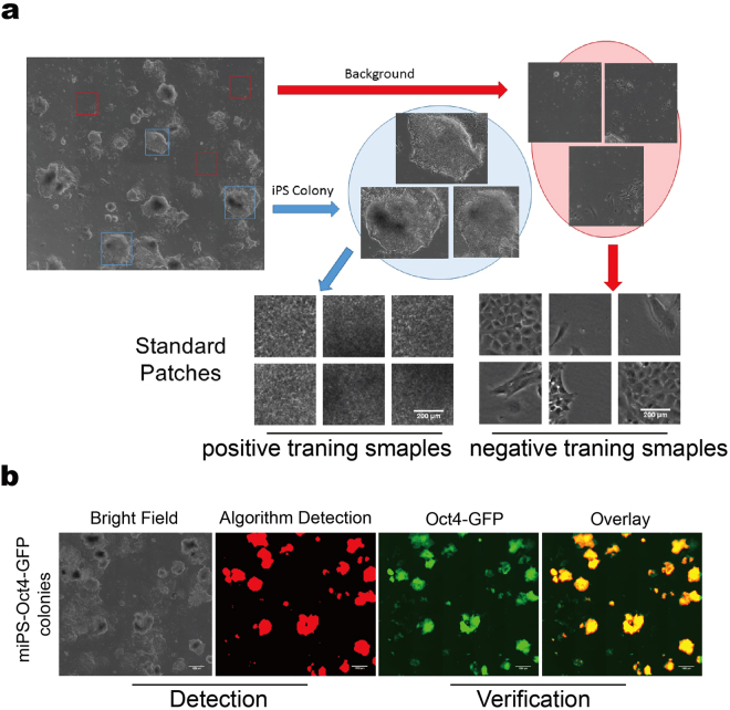
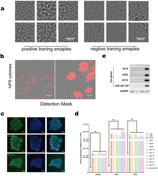
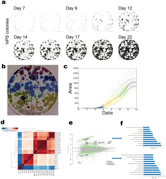
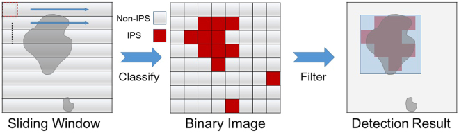
During cellular reprogramming, the mesenchymal-to-epithelial transition is accompanied by changes in morphology, which occur prior to iPSC colony formation. The current approach for detecting morphological changes associated with reprogramming purely relies on human experiences, which involve intensive amounts of upfront training, human error with limited quality control and batch-to-batch variations. Here, we report a time-lapse-based bright-field imaging analysis system that allows us to implement a label-free, non-invasive approach to measure morphological dynamics. To automatically analyse and determine iPSC colony formation, a machine learning-based classification, segmentation, and statistical modelling system was developed to guide colony selection. The system can detect and monitor the earliest cellular texture changes after the induction of reprogramming in human somatic cells on day 7 from the 20-24 day process. Moreover, after determining the reprogramming process and iPSC colony formation quantitatively, a mathematical model was developed to statistically predict the best iPSC selection phase independent of any other resources. All the computational detection and prediction experiments were evaluated using a validation dataset, and biological verification was performed. These algorithm-detected colonies show no significant differences (Pearson Coefficient) in terms of their biological features compared to the manually processed colonies using standard molecular approaches.


Conflict of interest statement

The authors declare that they have no competing interests.

Figures

Figure 1
Figure 1
Computer vision based detection of mice iPSC colonies. (a) Example of training patches. Positive samples cropped out as iPSC colonies and negative samples are MEF cells or background. (b) Verification of computer vision based method using the OCT4-GFP reporter. Panels from left to right indicate the bright field original image, a mask of computer detection, an OCT4-GFP fluorescent image, and an overlay between the fluorescence and binary images.
Figure 2
Figure 2
Computer vision based detection of human urinal cells (UCs) derived iPSC colonies. (a) Example of training patches. Positive samples are parts of iPSC colonies, and negative sample are other cells or background. Scale bar, 200μm. (b) A detection map. Example of iPSC colonies detection in one 6-well plate at Day 21 after reprogramming induction. Red regions indicate the position of iPSC colonies, which were detected by bright field image classification algorithm. A small area is enlarged to show the details of the detection. (c) Characterization of iPSCs, which were manually picked guided by the binary map. iPSC colonies (n = 3) were picked into cell culture plate based on the binary map. After 4–7 days culturing, iPSCs were stained with antibodies against known pluripotency surface markers (left). The nucleus was stained by DAPI (middle), merged images of two channel (right). Scale bar, 50μm. (d) Selected iPSCs were analysed for Nanog, SOX2 and OCT4 expression by qRT-PCR. Human ES cell line H1 was used as positive control. Error bars indicate Â ± s.d. of triplicates. P value is referenced to UCs. **Indicates P < 0.01. (e) PCR detection of exogenous eipsomal DNA in UC-iPSC. UC(Passage 3) and H1(Passage 46) cell were severed as the negative control, UC-iPSC(Passage21) was stably expanded, UC transiently transfected with eipsomal vector (pEP4EO2SET2K and pCEP4- miR-302-367 cluster) was served as the positive control.
Figure 3
Figure 3
Quantitative detection in synchronized time-lapse data for iPSCs selection prediction in human somatic cellular reprogramming. (a) A series detection map for the entire reprogramming process from day 7 to day 22. Binary indication masked out the colonies by classifier. (b) A segmented map. An example result of segmentation on a detection map, each segment represents a single colony (left); each colonies was individually registered for further process(right). (c) Growth curve of selected colony. Different colour indicted for different phases, which was calculated by the mathematical model; the purple colour stands for the mature phase. Solid line is an average of all training data and dash lines are some examples of training data. (d) Hierarchic clustering analysis of global gene expression based on Pearson correlation for Alg_iPSCs, H1 (C1–6) and Uri_C(1–2). (e) Differential gene expression profile between Alg_iPSCs (above) and UCs (below). The differentially expressed genes (red) are those with an adjusted P value 0.05 and fold change 3. (f) Functional annotations of genes differentially expressed between Alg_iPSCs (top) and UCs (bottom). Gene ontology (GO) was performed by DAVID, and enriched GO terms (biological processes) for each cell type are plotted with −log 10 of the adjusted P values.
Figure 4
Figure 4
The sliding window method was used to detect colonies from an unknown image. The small areas were divided into 96 × 96 pixel patches.

Similar articles

Cited by

References

    1. Wiley LA, et al. cGMP production of patient-specific iPSCs and photoreceptor precursor cells to treat retinal degenerative blindness. Sci Rep. 2016;6:30742. doi: 10.1038/srep30742. - DOI - PMC - PubMed
    1. Zhao T, et al. Humanized Mice Reveal Differential Immunogenicity of Cells Derived from Autologous Induced Pluripotent Stem Cells. Cell Stem Cell. 2015;17:353–359. doi: 10.1016/j.stem.2015.07.021. - DOI - PMC - PubMed
    1. Xue Y, et al. Generating a non-integrating human induced pluripotent stem cell bank from urine-derived cells. PLOS ONE. 2013;8:e70573. doi: 10.1371/journal.pone.0070573. - DOI - PMC - PubMed
    1. Zhou X, Cao X, Perlman Z, Wong ST. A computerized cellular imaging system for high content analysis in Monastrol suppressor screens. J Biomed Inform. 2006;39:115–125. doi: 10.1016/j.jbi.2005.05.008. - DOI - PubMed
    1. Wang M, Zhou X, King RW, Wong ST. Context based mixture model for cell phase identification in automated fluorescence microscopy. BMC Bioinformatics. 2007;8:32. doi: 10.1186/1471-2105-8-32. - DOI - PMC - PubMed

Publication types