

Susceptibility of different mouse strains to peri-implantitis
- PMID: 29044525
- PMCID: PMC5760471
- DOI: 10.1111/jre.12493
Susceptibility of different mouse strains to peri-implantitis
Abstract
Background and objective: Peri-implantitis (PI) is an inflammatory condition that affects the tissues surrounding dental implants. Although the pathogenesis of PI is not fully understood, evidence suggests that the etiology is multifactorial and may include a genetic component. The aim of this study was to investigate the role of genetics in the development of peri-implantitis.
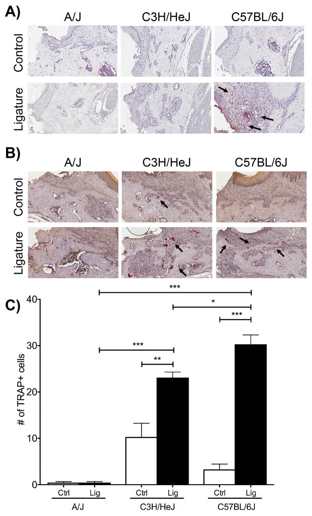
Material and methods: Four-week-old C57BL/6J, C3H/HeJ and A/J male mice had their left maxillary molars extracted. Implants were placed in the healed extraction sockets. Upon osseointegration, ligatures were placed around the implant head for 1 or 4 weeks to induce PI. Micro-computed tomography scanning was used to measure volumetric bone loss. Histological analyses were also performed to evaluate collagen organization and the presence of neutrophils and osteoclasts.
Results: Radiographically, comparing the ligature-treated mice, C57BL/6J displayed the greatest amount of bone loss, followed by C3H/HeJ and A/J mice at 1 and 4 weeks. Histologically, at 1 week, C57BL/6J mice presented with the highest numbers of neutrophils and osteoclasts. At 4 weeks, C57BL/6J mice presented with the most active bone remodeling compared with the other two strains.
Conclusion: There were significant differences in the severity of peri-implantitis among the different mouse strains, suggesting that the genetic framework can affect implant survival and success. Future work is needed to dissect the genetic contribution to the development of peri-implantitis.
Keywords: bone loss; dental implant; genetics; murine.
© 2017 John Wiley & Sons A/S. Published by John Wiley & Sons Ltd.
Figures


Similar articles
-
Role of toll-like receptor 2 in inflammation and alveolar bone loss in experimental peri-implantitis versus periodontitis.J Periodontal Res. 2018 Feb;53(1):98-106. doi: 10.1111/jre.12492. Epub 2017 Sep 5. J Periodontal Res. 2018. Retraction in: J Periodontal Res. 2021 Jan;56(1):203. doi: 10.1111/jre.12815. PMID: 28872184 Free PMC article. Retracted.
-
Comparing the Healing Potential of Late-Stage Periodontitis and Peri-Implantitis.J Oral Implantol. 2017 Dec;43(6):437-445. doi: 10.1563/aaid-joi-D-17-00157. Epub 2017 Oct 24. J Oral Implantol. 2017. PMID: 29064761
-
Evaluation of Bone Loss in Implants Adjacent to a Tooth or Edentulous Area in Peri-Implantitis and Control Murine Models.J Oral Implantol. 2025 Feb 1;51(1):98-104. doi: 10.1563/aaid-joi-D-24-00100. J Oral Implantol. 2025. PMID: 39731390
-
Basis of bone metabolism around dental implants during osseointegration and peri-implant bone loss.J Biomed Mater Res A. 2017 Jul;105(7):2075-2089. doi: 10.1002/jbm.a.36060. Epub 2017 Mar 28. J Biomed Mater Res A. 2017. PMID: 28281321 Review.
-
"Peri-Implantitis": A Complication of a Foreign Body or a Man-Made "Disease". Facts and Fiction.Clin Implant Dent Relat Res. 2016 Aug;18(4):840-9. doi: 10.1111/cid.12427. Epub 2016 May 30. Clin Implant Dent Relat Res. 2016. PMID: 27238274 Review.
Cited by
-
Revealing leukocyte populations in human peri-implantitis and periodontitis using flow cytometry.Clin Oral Investig. 2023 Sep;27(9):5499-5508. doi: 10.1007/s00784-023-05168-y. Epub 2023 Jul 25. Clin Oral Investig. 2023. PMID: 37490117
-
Peri-implant inflammation increases the risk of osteonecrosis in mice treated with bisphosphonate.J Periodontol. 2025 Apr 15:10.1002/JPER.24-0760. doi: 10.1002/JPER.24-0760. Online ahead of print. J Periodontol. 2025. PMID: 40231895
-
Peri-Implant Surgical Treatment Downregulates the Expression of sTREM-1 and MMP-8 in Patients with Peri-Implantitis: A Prospective Study.Int J Environ Res Public Health. 2022 Mar 18;19(6):3627. doi: 10.3390/ijerph19063627. Int J Environ Res Public Health. 2022. PMID: 35329310 Free PMC article.
-
Correlations of FCGR2A 131R/H and FCGR3A 158V/F Polymorphisms with the Susceptibility of Peri-implantitis in Chinese Han Population.Mol Biotechnol. 2025 Jun;67(6):2254-2261. doi: 10.1007/s12033-024-01193-8. Epub 2024 May 21. Mol Biotechnol. 2025. PMID: 38771420
-
Temporal changes of periodontal tissue pathology in a periodontitis animal model.J Periodontal Implant Sci. 2023 Aug;53(4):248-258. doi: 10.5051/jpis.2203420171. Epub 2022 Nov 23. J Periodontal Implant Sci. 2023. PMID: 36468486 Free PMC article.
References
-
- Mombelli A, Lang NP. The diagnosis and treatment of peri-implantitis. Periodontology 2000. 1998;17:63–76. - PubMed
-
- Derks J, Schaller D, Hakansson J, Wennstrom JL, Tomasi C, Berglundh T. Effectiveness of Implant Therapy Analyzed in a Swedish Population: Prevalence of Peri-implantitis. Journal of dental research. 2016;95:43–49. - PubMed
-
- Sullivan RM. Implant dentistry and the concept of osseointegration: a historical perspective. Journal of the California Dental Association. 2001;29:737–745. - PubMed
-
- Heitz-Mayfield LJ, Lang NP. Comparative biology of chronic and aggressive periodontitis vs. peri-implantitis. Periodontology 2000. 2010;53:167–181. - PubMed
MeSH terms
Grants and funding
LinkOut - more resources
Full Text Sources
Other Literature Sources
Research Materials
Miscellaneous
