. 2018 Jan;36(1):65-78.
doi: 10.1002/stem.2722. Epub 2017 Oct 31.

Neuroprotective Effects of Human Mesenchymal Stem Cells and Platelet-Derived Growth Factor on Human Retinal Ganglion Cells

Affiliations

Neuroprotective Effects of Human Mesenchymal Stem Cells and Platelet-Derived Growth Factor on Human Retinal Ganglion Cells

Andrew Osborne et al. Stem Cells. 2018 Jan.

Abstract

Optic neuropathies such as glaucoma occur when retinal ganglion cells (RGCs) in the eye are injured. Strong evidence suggests mesenchymal stem cells (MSCs) could be a potential therapy to protect RGCs; however, little is known regarding their effect on the human retina. We, therefore, investigated if human MSCs (hMSCs), or platelet-derived growth factor (PDGF) as produced by hMSC, could delay RGC death in a human retinal explant model of optic nerve injury. Our results showed hMSCs and the secreted growth factor PDGF-AB could substantially reduce human RGC loss and apoptosis following axotomy. The neuroprotective pathways AKT, ERK, and STAT3 were activated in the retina shortly after treatments with labeling seen in the RGC layer. A dose dependent protective effect of PDGF-AB was observed in human retinal explants but protection was not as substantial as that achieved by culturing hMSCs on the retina surface which resulted in RGC cell counts similar to those immediately post dissection. These results demonstrate that hMSCs and PDGF have strong neuroprotective action on human RGCs and may offer a translatable, therapeutic strategy to reduce degenerative visual loss. Stem Cells 2018;36:65-78.

Keywords: Degeneration; Human; Mesenchymal stem cells; Neuroprotection; Retina.


Figures

Figure 1
Figure 1
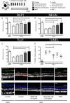
Human RGC neuroprotection by PDGF or human MSCs following regular medium replenishment. Human retinal explants were cultured for 7 days in medium containing PDGF or with the addition of hMSCs pipetted directly onto the RGC layer surface. (A–C): Neuronal survival in the RGC layer was quantified immediately post dissection, 0DEV or after 7 days’ culture (7DEV) (n = 6). (D): Number of apoptotic, NeuN+ cells were quantified and expressed as a percentage of all NeuN+ cells in the RGC layer (n = 6). (E, F): Representative images of RGCs (NeuN ‐ green or TUJ1 ‐ blue) in human retinal explants with apoptotic cells labeled red. ×20 objective, scale bar = 50 µm, *, p < .05; **, p < .01; ***, p < .001. Bar and scatter graphs show mean ± s.e.m with (n) number indicating the number of unique post‐mortem eyes used. The schematic in the top left shows the number of explants processed from each retina and the treatment time course. Abbreviations: 0DEV, 0 days ex vivo; 7DEV, 7 days ex vivo; MSC, mesenchymal stem cell; PDGF, platelet‐derived growth factor; RGCL, retinal ganglion cell layer.
Figure 2
Figure 2
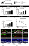
Human RGC neuroprotection after a single treatment of PDGF or human MSCs. Human retinal explants were cultured for 7 days in medium containing PDGF or with the addition of hMSCs pipetted directly onto the RGC layer surface. (A–C): Neuronal survival in the RGC layer was quantified immediately post dissection, 0DEV or after 7 days’ culture with no further medium change (7DEV) (n = 6) (D): Number of apoptotic, NeuN+ cells were quantified and expressed as a percentage of all NeuN+ cells in the RGC layer (n = 6). (E, F): Magnified, representative images of RGCs (NeuN ‐ green or TUJ1 ‐ blue) in human retinal explants with apoptotic cells labeled red. ×40 objective, scale bar = 50 µm, *, p < .05, **, p < .01 and ***, p < .001. Bar and scatter graphs show mean ± s.e.m with (n) number indicating the number of unique post‐mortem eyes used. The schematic in the top left shows the number of explants processed from each retina and the treatment time course. Abbreviations: 0DEV, 0 days ex vivo; 7DEV, 7 days ex vivo; MSC, mesenchymal stem cell; PDGF, platelet‐derived growth factor; RGCL, retinal ganglion cell layer.
Figure 3
Figure 3
Necrotic cell damage in human retinal explants over time and the detectable levels of PDGF in the culture medium. (A): Lactate dehydrogenase within the culture medium relative to the protein content within human retinal explants treated with PDGF or hMSCs (n = 6), *, p < .05. (B): Detection of PDGF‐AB in the culture medium post addition at 0DEV (n = 6), *, p < .05 compared with 0DEV PDGF‐AB (150 ng/ml), Ω p < .05 compared with 0DEV PDGF‐AB (50 ng/ml). Bar graphs show mean ± s.e.m with (n) number indicating the number of unique post‐mortem eyes used. The schematic in the top left shows the number of explants processed from each retina and the treatment time course. Abbreviations: DEV, days ex vivo; MSC, mesenchymal stem cell; PDGF, platelet‐derived growth factor.
Figure 4
Figure 4
Activation of downstream signaling pathways in human retinal explants after a single treatment with PDGF or human MSCs. (A–D): Activation of cell survival pathways 1 and 3 days after human explant culture in PDGF supplemented medium or addition of hMSCs relative to untreated, same time point controls (n = 4), *, p < .05. (E): Pro‐apoptotic BAX expression in treatment groups relative to untreated, same time point controls (n = 4), *, p < .05. Bar graphs show mean ± s.e.m with (n) number indicating the number of unique post‐mortem eyes used. The schematic in the top left shows the number of explants processed from each retina and the treatment time course. Abbreviations: DEV, days ex vivo; MSC, mesenchymal stem cell; PDGF, platelet‐derived growth factor.
Figure 5
Figure 5
PDGFR activation and downstream survival signaling could be seen within the retina and RGCL after PDGF and human MSC treatments. (A): Increased PDGFRα signaling (purple) was observed 3 days after PDGF‐AB or hMSC treatments with elevation in phosphorylated STAT3 (p‐STAT3 ‐ yellow) and phosphorylated S6 (p‐S6 ‐ teal) signaling in the RGCL. PDGFRβ activation (purple) was detected on RGCL cells and colocalized with activated ERK (p‐ERK ‐ yellow) or p‐S6 (teal). ×40 objective, scale bar = 50 µm, arrows show colocalization in the RGCL. Representative images from three experimental repeats. Abbreviations: DEV, days ex vivo; MSC, mesenchymal stem cell; PDGFR, platelet‐derived growth factor receptor; RGCL, retinal ganglion cell layer.
Figure 6
Figure 6
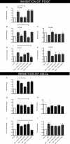
The beneficial effects of PDGF or human MSCs could be reduced using a variety of inhibitors when assessed 3 days after treatment. (A–E): PDGF in combination with a PDGF inhibitor no longer activated survival pathways or protected against a decrease in retinal ganglion cell (TUJ1) expression (n = 4). Inhibitors of PI3K, ERK, or STAT3 effectively blocked individual pathway signaling but did not improve retinal (BAX) or retinal ganglion cell specific (TUJ1) survival (n = 4). (F–J): hMSCs could still offer some protection even in the presence of inhibitors against PDGF, PI3K, ERK, or STAT3 (n = 4), *, p < .05; **, p < .01. Bar graphs show mean ± s.e.m with (n) number indicating the number of unique post‐mortem eyes used. Abbreviations: MSC, mesenchymal stem cell; PDGF, platelet‐derived growth factor.
Figure 7
Figure 7
Inhibition of PDGF in human retinal explants cultured with human MSCs did not prevent MSC mediated neuroprotection. (A–C): Neuronal survival in the RGCL was quantified immediately post dissection, 0DEV or after 7 days’ culture with no further medium change (7DEV) (n = 3). (D): Number of apoptotic, NeuN+ cells were quantified and expressed as a percentage of all NeuN+ cells in the RGC layer (n = 3). (E): Human MSCs (CD105+ ‐ green and CD73+ ‐ red cells) could be visualized on the surface of the human retina in close proximity to the RGC layer. (F): Human MSCs remained viable throughout the 7‐day culture with little evidence of apoptotic labeling (red staining on green cells). (G): Multiple growth factors and neurotrophins could be measured in the hMSC secretome after 72 hours in culture. ×40 objective, scale bar = 50 µm. Bar and scatter graphs show mean ± s.e.m with (n) number indicating the number of unique post‐mortem eyes used. The schematic in the top left shows the number of explants processed from each retina and the treatment time course. Abbreviations: 0DEV, 0 days’ ex vivo; 7DEV, 7 days ex vivo; Ang, angiopoietin; EGF, epidermal growth factor; FGF, fibroblast growth factor; HGF, hepatocyte growth factor; IL, interleukin; MSC, mesenchymal stem cell; MCP, monocyte chemotactic protein; PDGF, platelet‐derived growth factor; RGCL, retinal ganglion cell layer; VEGF, vascular endothelial growth factor.

References

    1. Yu S, Tanabe T, Dezawa M et al. Effects of bone marrow stromal cell injection in an experimental glaucoma model. Biochem Biophys Res Commun 2006;344:1071–1079. - PubMed
    1. Zwart I, Hill A, Al‐Allaf F et al. Umbilical cord blood mesenchymal stromal cells are neuroprotective and promote regeneration in a rat optic tract model. Exp Neurol 2009;216:439–448. - PubMed
    1. Johnson T, Bull N, Hunt D et al. Neuroprotective effects of intravitreal mesenchymal stem cell transplantation in experimental glaucoma. Invest Ophthalmol Vis Sci 2010;51:2051–2059. - PMC - PubMed
    1. Hu Y, Tan H, Wang X et al. Bone marrow mesenchymal stem cells protect against retinal ganglion cell loss in aged rats with glaucoma. Clin Interv Aging 2013;8:1467–1470. - PMC - PubMed
    1. Mesentier‐Louro L, Zaverucha‐do‐Valle C, da Silva‐Junior A et al. Distribution of mesenchymal stem cells and effects on neuronal survival and axon regeneration after optic nerve crush and cell therapy. PLoS One 2014;9:e110722. - PMC - PubMed

Publication types

MeSH terms

Substances