

Tumor radiomic heterogeneity: Multiparametric functional imaging to characterize variability and predict response following cervical cancer radiation therapy
- PMID: 29044908
- PMCID: PMC5899626
- DOI: 10.1002/jmri.25874
Tumor radiomic heterogeneity: Multiparametric functional imaging to characterize variability and predict response following cervical cancer radiation therapy
Abstract
Background: Robust approaches to quantify tumor heterogeneity are needed to provide early decision support for precise individualized therapy.
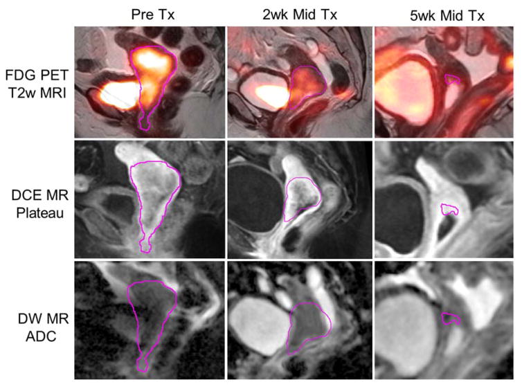
Purpose: To conduct a technical exploration of longitudinal changes in tumor heterogeneity patterns on dynamic contrast-enhanced (DCE) magnetic resonance imaging (MRI), diffusion-weighted imaging (DWI) and FDG positron emission tomography / computed tomography (PET/CT), and their association to radiation therapy (RT) response in cervical cancer.
Study type: Prospective observational study with longitudinal MRI and PET/CT pre-RT, early-RT (2 weeks), and mid-RT (5 weeks).
Population: Twenty-one FIGO IB2 -IVA cervical cancer patients receiving definitive external beam RT and brachytherapy.
Field strength/sequence: 1.5T, precontrast axial T1 -weighted, axial and sagittal T2 -weighted, sagittal DWI (multi-b values), sagittal DCE MRI (<10 sec temporal resolution), postcontrast axial T1 -weighted.
Assessment: Response assessment 1 month after completion of treatment by a board-certified radiation oncologist from manually delineated tumor volume changes.
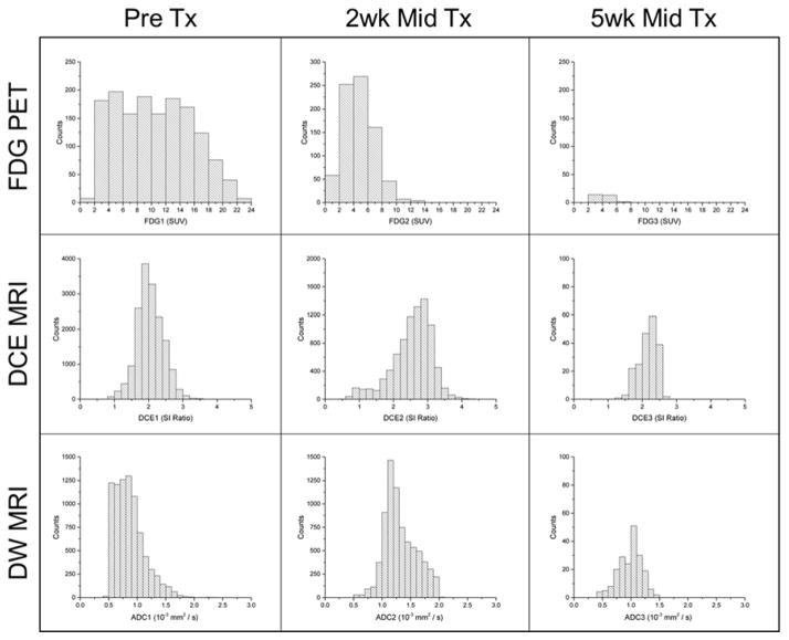
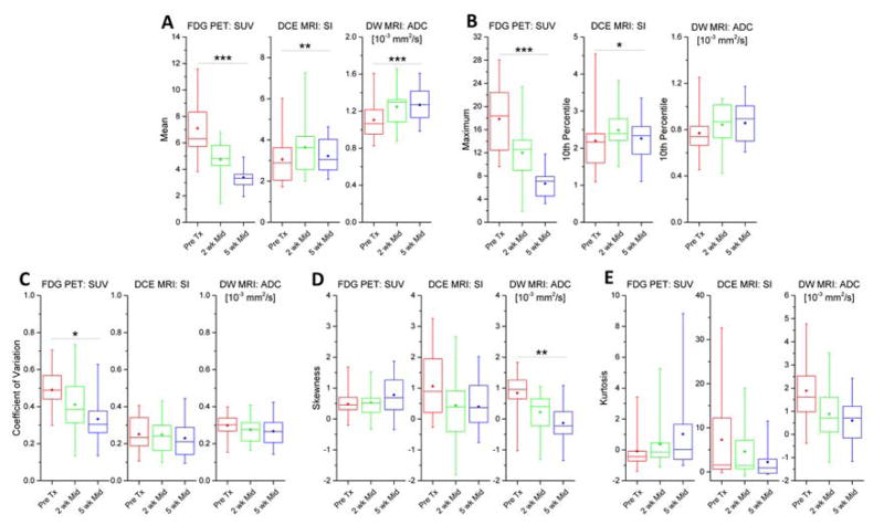
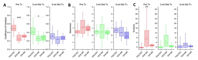
Statistical tests: Intensity histogram (IH) quantiles (DCE SI10% and DWI ADC10% , FDG-PET SUVmax ) and distribution moments (mean, variance, skewness, kurtosis) were extracted. Differences in IH features between timepoints and modalities were evaluated by Skillings-Mack tests with Holm's correction. Area under receiver-operating characteristic curve (AUC) and Mann-Whitney testing was performed to discriminate treatment response using IH features.
Results: Tumor IH means and quantiles varied significantly during RT (SUVmean : ↓28-47%, SUVmax : ↓30-59%, SImean : ↑8-30%, SI10% : ↑8-19%, ADCmean : ↑16%, P < 0.02 for each). Among IH heterogeneity features, FDG-PET SUVCoV (↓16-30%, P = 0.011) and DW-MRI ADCskewness decreased (P = 0.001). FDG-PET SUVCoV was higher than DCE-MRI SICoV and DW-MRI ADCCoV at baseline (P < 0.001) and 2 weeks (P = 0.010). FDG-PET SUVkurtosis was lower than DCE-MRI SIkurtosis and DW-MRI ADCkurtosis at baseline (P = 0.001). Some IH features appeared to associate with favorable tumor response, including large early RT changes in DW-MRI ADCskewness (AUC = 0.86).
Data conclusion: Preliminary findings show tumor heterogeneity was variable between patients, modalities, and timepoints. Radiomic assessment of changing tumor heterogeneity has the potential to personalize treatment and power outcome prediction.
Level of evidence: 2 Technical Efficacy: Stage 3 J. Magn. Reson. Imaging 2018;47:1388-1396.
Keywords: DCE; DWI; MRI; PET; radiomics; tumor heterogeneity.
© 2017 International Society for Magnetic Resonance in Medicine.
Conflict of interest statement
Stephen R. Bowen was supported by a Research Scholar Award from the Radiological Society of North American and an NIH/NCI grant for work performed as part of the current study. William T.C. Yuh, Daniel S. Hippe, Savannah C. Partridge, Michael V. Knopp, and Nina A. Mayr were all suported by an NIH/NCI grant for work performed as part of the current study. Dennis Nelson has a commercial interest as President of MIM Software, Inc. for work performed outside of the current study. Paul Kinahan has a commercial interest as co-founder of PET/X, LLC for work performed outside of the current study. Saba Elias, Guang Jia, Zhibin Huang, Norman J. Beauchamp, George A. Sandison, and Simon S. Lo declare no conflicts of interest.
Figures
Similar articles
-
A prospective study of DWI, DCE-MRI and FDG PET imaging for target delineation in brachytherapy for cervical cancer.Radiother Oncol. 2016 Sep;120(3):519-525. doi: 10.1016/j.radonc.2016.08.002. Epub 2016 Aug 12. Radiother Oncol. 2016. PMID: 27528120
-
Pre-treatment prediction of early response to chemoradiotherapy by quantitative analysis of baseline staging FDG-PET/CT and MRI in locally advanced cervical cancer.Acta Radiol. 2021 Jul;62(7):940-948. doi: 10.1177/0284185120943046. Epub 2020 Jul 28. Acta Radiol. 2021. PMID: 32722967
-
Variability of target and normal structure delineation using multimodality imaging for radiation therapy of pancreatic cancer.Int J Radiat Oncol Biol Phys. 2014 Jul 1;89(3):633-40. doi: 10.1016/j.ijrobp.2014.02.035. Epub 2014 Apr 20. Int J Radiat Oncol Biol Phys. 2014. PMID: 24755533
-
A Comprehensive Comparison of CT, MRI, Positron Emission Tomography or Positron Emission Tomography/CT, and Diffusion Weighted Imaging-MRI for Detecting the Lymph Nodes Metastases in Patients with Cervical Cancer: A Meta-Analysis Based on 67 Studies.Gynecol Obstet Invest. 2017;82(3):209-222. doi: 10.1159/000456006. Epub 2017 Feb 10. Gynecol Obstet Invest. 2017. PMID: 28183074 Review.
-
Update of the role of PET/CT and PET/MRI in the management of patients with cervical cancer.Hell J Nucl Med. 2016 Sep-Dec;19(3):254-268. doi: 10.1967/s002449910409. Epub 2016 Nov 8. Hell J Nucl Med. 2016. PMID: 27824966 Review.
Cited by
-
Radiomics-based prediction of two-year clinical outcome in locally advanced cervical cancer patients undergoing neoadjuvant chemoradiotherapy.Radiol Med. 2022 May;127(5):498-506. doi: 10.1007/s11547-022-01482-9. Epub 2022 Mar 24. Radiol Med. 2022. PMID: 35325372 Free PMC article. Review.
-
The transformational potential of molecular radiomics.J Med Radiat Sci. 2023 Apr;70 Suppl 2(Suppl 2):77-88. doi: 10.1002/jmrs.626. Epub 2022 Oct 13. J Med Radiat Sci. 2023. PMID: 36238997 Free PMC article. Review.
-
Prediction of out-of-field recurrence after chemoradiotherapy for cervical cancer using a combination model of clinical parameters and magnetic resonance imaging radiomics: a multi-institutional study of the Japanese Radiation Oncology Study Group.J Radiat Res. 2022 Jan 20;63(1):98-106. doi: 10.1093/jrr/rrab104. J Radiat Res. 2022. PMID: 34865079 Free PMC article.
-
Radiomics Based on Contrast-Enhanced CT for Recognizing c-Met-Positive Hepatocellular Carcinoma: a Noninvasive Approach to Predict the Outcome of Sorafenib Resistance.Mol Imaging Biol. 2023 Dec;25(6):1073-1083. doi: 10.1007/s11307-023-01870-1. Epub 2023 Nov 6. Mol Imaging Biol. 2023. PMID: 37932610
-
A decade of multi-modality PET and MR imaging in abdominal oncology.Br J Radiol. 2021 Oct 1;94(1126):20201351. doi: 10.1259/bjr.20201351. Epub 2021 Aug 13. Br J Radiol. 2021. PMID: 34387508 Free PMC article. Review.
References
-
- Hanahan D, Weinberg RA. Hallmarks of cancer: the next generation. Cell. 2011;144:646–74. - PubMed
-
- Bachtiary B, Boutros PC, Pintilie M. Gene expression profiling in cervical cancer: an exploration of intratumor heterogeneity. Clin Cancer Res. 2006;12:5632–40. - PubMed
-
- Ellingsen C, Natvig I, Gaustad JV, Gulliksrud K, Egeland TA, Rofstad EK. Human cervical carcinoma xenograft models for studies of the physiological microenvironment of tumors. J Cancer Res Clin Oncol. 2009;135:1177–84. - PubMed
Publication types
MeSH terms
Substances
Grants and funding
LinkOut - more resources
Full Text Sources
Other Literature Sources
Medical
Research Materials
