. 2018 Jan;19(1):36-43.
doi: 10.1111/tra.12536. Epub 2017 Dec 1.

Recruitment of 7SL RNA to assembling HIV-1 virus-like particles

Affiliations

Recruitment of 7SL RNA to assembling HIV-1 virus-like particles

Michelle S Itano et al. Traffic. 2018 Jan.

Abstract

Retroviruses incorporate specific host cell RNAs into virions. In particular, the host noncoding 7SL RNA is highly abundant in all examined retroviruses compared with its cellular levels or relative to common mRNAs such as actin. Using live cell imaging techniques, we have determined that the 7SL RNA does not arrive with the HIV-1 RNA genome. Instead, it is recruited contemporaneously with assembly of the protein HIV-1 Gag at the plasma membrane. Further, we demonstrate that complexes of 7SL RNA and Gag can be immunoprecipitated from both cytosolic and plasma membrane fractions. This indicates that 7SL RNAs likely interact with Gag prior to high-order Gag multimerization at the plasma membrane. Thus, the interactions between Gag and the host RNA 7SL occur independent of the interactions between Gag and the host endosomal sorting complex required for transport (ESCRT) proteins, which are recruited temporarily at late stages of assembly. The interactions of 7SL and Gag are also independent of interactions of Gag and the HIV-1 genome which are seen on the plasma membrane prior to assembly of Gag.

Keywords: RNA; cytoplasm; diffusion; fluorescence microscopy; noncoding RNAs; retrovirus; transport.


Conflict of interest statement

The authors declare no conflict of interest.

Figures

Fig 1.
Fig 1.. Strategy for imaging 7SL RNA.
A) 7SL contains an Alu domain needed for saturable uptake and an S domain. SRP19 binding sites are shaded. The sequence of 7SL1 is shown, and the nucleotide changes in 7SL2 are in red. B) To create 7SL-MS2, two S domain stem loops were replaced with MS2 stem loops (blue). The sequence and predicted structures of 7SL-MS2 RNAs are shown.
Fig 2.
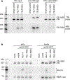
Fig 2.. 7SL-MS2 binds stably to MCP-mCherry and competes for packaging with endogenous 7SL.
A) After transfecting HeLa cells stably expressing MCP-NLS-mCherry with either empty vector or plasmids expressing 7SL1-MS2 or 7SL2-MS2, the resulting cell lysates were subjected to immunoprecipitation with anti-mCherry antibodies. RNAs in the starting lysates, immunoprecipitates, and supernatants were subjected to Northern blotting to detect MS2-stem loop containing RNAs (bottom) or all 7SL RNAs (top).B) HEK293T cells were transfected with pHIVpuro and either pAlu114, p7SL1-MS2, p7SL2-MS2 or empty vector. After 48 hours, virus was harvested from the media, and normalized by Western blotting for Gag. RNAs extracted from cells and virions were subjected to Northern analysis to detect 7SL RNAs (top), Alu114 (middle) and tRNA-Lys3 (bottom panel).
Fig 3.
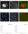
Fig 3.. Visualization of individual 7SL RNA.
A) HeLa MCP-NLS-mCherry cells were transfected with Gag/Gag-mEGFP (5:1), and 7SL1-MS2 and observed with epifluorescence illumination following deconvolution after 26 h where we visualize 7SL-MS2 puncta outside of the nucleus when Gag is expressed. Scale bar, 4 μm. B) Virus like particles isolated from the supernatant of 293T cells transfected 7SL2-MS2, MCP -NLS-mCherry and proviral NL4–3_SAFE and NL4–3_MA-YFP_SAFE (5:1 ratio) and imaged in TIR-FM, a gaussian filter (sigma = 1) was used on the red image). Blue arrows indicate Gag and 7SL-MS2 puncta that colocalize. Scale bar, 2 μm. C,D) Graphs of the total percentage of 7SL-MS2 fluorescence signal that colocalizes with Gag signal in the deconvolved image in (A) or the VLP image in (B), C and D, respectively.
Fig 4.
Fig 4.. 7SL1-MS2 co-assembles with Gag.
293T cells were transiently transfected with MCP-NLS-mCherry, 7SL1-MS2 and proviral NL4–3_SAFE and NL4–3_MA-YFP_SAFE (5:1 ratio), and were observed with TIR-FM after 5 h. A) Images of an assembling punctum containing both labeled Gag and RNA. Elapsed time is in seconds. B) Plots of normalized fluorescence intensity for the labeled RNA and Gag-MA-YFP signals for the VLP shown in (A). Scale bars, 500 nm.
Fig 5.
Fig 5.. 7SL associates with Gag-GFP in the cytoplasm and at the plasma membrane.
(A). HEK293T cells were transfected with Gag-GFP and cells harvested after 24 hours. After removing nuclei and unbroken cells, lysates were fractionated in flotation gradients. Proteins from gradient fractions were subjected to Western blotting to detect Gag (top panel), while extracted RNAs were subjected to Northern blotting to detect 7SL RNA (bottom panel). Gag-GFP was predominantly in Fractions 2, 3 (plasma membrane fractions) and 10 (cytoplasm). 7SL was primarily in fractions 3 and 9–11. (B). Fractions 2, 3 and 10 were subjected to immunoprecipitation with anti-GFP antibodies. RNAs extracted from the input, the supernatants and the immunoprecipitate were subjected to Northern blotting to detect 7SL RNA. The top panel is a light exposure, the bottom, a much darker one. 7SL is detected in the immunoprecipitates from both fraction 3 (plasma membrane) and fraction 10 (cytoplasm).

Similar articles

Cited by

References

    1. Bishop JM, Levinson WE, Sullivan D, Fanshier L, Quintrell N, Jackson J. The low molecular weight RNAs of Rous sarcoma virus. II. The 7 S RNA. Virology. 1970;42(4):927–937. - PubMed
    1. Faras AJ, Garapin AC, Levinson WE, Bishop JM, Goodman HM. Characterization of the low-molecular-weight RNAs associated with the 70S RNA of Rous sarcoma virus. J Virol. 1973;12(2):334–342. - PMC - PubMed
    1. Bishop JM, Levinson WE, Quintrell N, Sullivan D, Fanshier L, Jackson J. The low molecular weight RNAs of Rous sarcoma virus. I. The 4 S RNA. Virology. 1970;42(1):182–195. - PubMed
    1. Erikson E, Erikson RL, Henry B, Pace NR. Comparison of oligonucleotides produced by RNase T1 digestion of 7 S RNA from avian and murine oncornaviruses and from uninfected cells. Virology. 1973;53(1):40–46. - PubMed
    1. Sawyer RC, Dahlberg JE. Small RNAs of Rous sarcoma virus: characterization by two-dimensional polyacrylamide gel electrophoresis and fingerprint analysis. J Virol. 1973;12(6):1226–1237. - PMC - PubMed

Publication types

MeSH terms

Substances

LinkOut - more resources