. 2018 Feb;22(2):904-912.
doi: 10.1111/jcmm.13378. Epub 2017 Oct 17.

Aquaporin-1 inhibition reduces metastatic formation in a mouse model of melanoma

Affiliations

Aquaporin-1 inhibition reduces metastatic formation in a mouse model of melanoma

Laura Simone et al. J Cell Mol Med. 2018 Feb.

Abstract

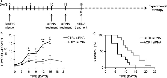
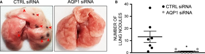
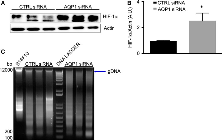
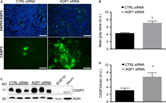
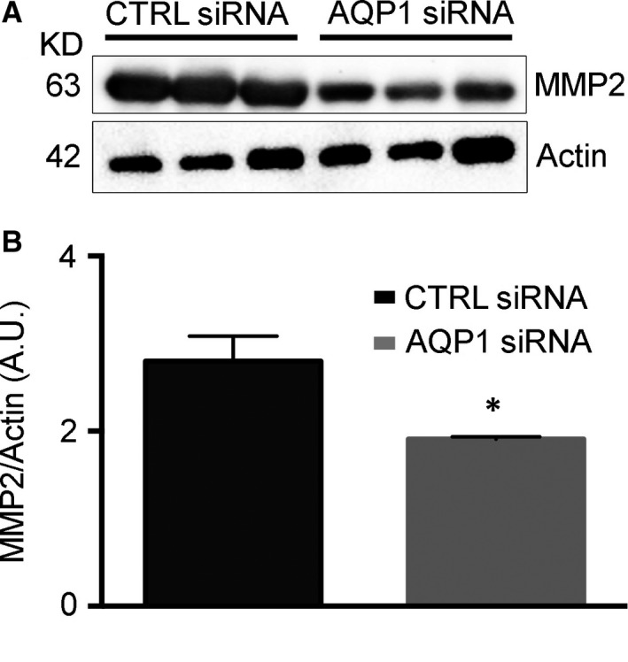
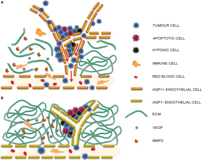
Aquaporin-1 (AQP1) is a proangiogenic water channel protein promoting endothelial cell migration. We previously reported that AQP1 silencing by RNA interference reduces angiogenesis-dependent primary tumour growth in a mouse model of melanoma. In this study, we tested the hypothesis that AQP1 inhibition also affects animal survival and lung nodule formation. Melanoma was induced by injecting B16F10 cells into the back of C57BL6J mice. Intratumoural injection of AQP1 siRNA and CTRL siRNA was performed 10 days after tumour cell implantation. Lung nodule formation was analysed after the death of the mice. Western blot was used to quantify HIF-1α, caspase-3 (CASP3) and metalloproteinase-2 (MMP2) protein levels. We found that AQP1 knock-down (KD) strongly inhibited metastatic lung nodule formation. Moreover, AQP1 siRNA-treated mice showed a twofold survival advantage compared to mice receiving CTRL siRNAs. The reduced AQP1-dependent tumour angiogenesis caused a hypoxic condition, evaluated by HIF-1α significant increase, in turn causing an increased level of apoptosis in AQP1 KD tumours, assessed by CASP3 quantification and DNA fragmentation. Importantly, a decreased level of MMP2 after AQP1 KD indicated a decreased activity against extracellular matrix associated with reduced vascularization and metastatic formation. In conclusion, these findings highlight an additional role for AQP1 as an important determinant of tumour dissemination by facilitating tumour cell extravasation and metastatic formation. This study adds knowledge on the role played by AQP1 in tumour biology and supports the view of AQP1 as a potential drug target for cancer therapy.

Keywords: antiangiogenic therapy; apoptosis; aquaporin-1; endothelial migration; extracellular matrix; melanoma; metastasis; tumour angiogenesis.


Figures

Figure 1
Figure 1
AQP1 silencing prolongs survival in the mouse model of melanoma. (A) Schematic representation of mouse treatment strategy for the analysis of tumour growth and animal survival. (B) Effect of AQP1 silencing on subcutaneous tumour growth. The diagram shows tumour growth of CTRL group (black line, n = 8) versus AQP1 siRNA‐treated group (grey line, n = 8). Data are expressed as the means ± S.E.M. (*P < 0.05, **P < 0.005). (C) Kaplan–Meier plot of CTRL siRNA‐ (black line, n = 12) versus AQP1 siRNA‐treated group (grey line, n = 13).
Figure 2
Figure 2
AQP1 silencing impairs metastatic lung nodule formation. (A) Representative images of lungs from CTRL and AQP1 siRNA‐treated mice after subcutaneous injection of B16F10. (B) Number of metastases counted at the day of death (in the x‐axis) in the lung from CTRL (n = 7) and AQP1 (n = 8) siRNA‐treated mice. Data are expressed as the means ± S.E.M. (*P < 0.05).
Figure 3
Figure 3
AQP1 silencing causes hypoxia and DNA degradation. (A) Immunoblot analysis of HIF‐1α expression in tumour explanted from CTRL siRNA‐ and AQP1 siRNA‐treated mice, as indicated. Actin was used as internal control for protein concentration. (B) Bar chart showing the densitometric analysis of HIF‐1α expression in CTRL (n = 3) and AQP1 (n = 3) siRNA‐treated mice. Data are expressed as the means ± S.E.M. (*P < 0.05). (C) Agarose gel electrophoresis showing the integrity of genomic DNA (gDNA, arrow) extract from tumours of CTRLs (n = 3) and AQP1 (n = 3) siRNA‐treated mice. Lane DNA ladder contains a 100‐bp marker.
Figure 4
Figure 4
AQP1 silencing causes increased caspase activity. (A) Immunofluorescence analysis performed on tumour cryosections with antibodies against CASP3 (green) at lower magnification (top, scale bar 100 μm) and with DAPI staining of nuclei (blue) and at higher magnification (bottom, scale bar 20 μm). (B) Quantitative analysis of immunofluorescence experiments shown in (A). The bar chart shows the fluorescence intensity of CASP3 staining performed on whole images converted in grey scale. Data are expressed as the means ± S.E.M. (*P < 0.05). (C) Immunoblot analysis of cleaved CASP3 expression in tumours explanted from CTRL siRNA‐ and AQP1 siRNA‐treated mice, as indicated. Actin was used as internal control for protein concentration. B16F10 cells treated with 2 μM of staurosporine (stauro) for 48 hrs were used as positive control for CASP3 activation; untreated B16F10 cells were used as negative control. (D) Bar chart showing the densitometry analysis of CASP3 expression in CTRL (n = 5) and AQP1 (n = 5) siRNA‐treated mice. Data are expressed as the means ± S.E.M. (*P < 0.05).
Figure 5
Figure 5
Metalloproteinase‐2 expression is reduced in AQP1 siRNA‐treated tumours. (A) Western blot analysis revealing a reduced MMP2 expression in AQP1 siRNA‐compared with CTRL siRNA‐treated tumours. Actin was used as internal control for protein concentration. (B) Bar chart showing the densitometric analysis of MMP2 expression in CTRL (n = 5) and AQP1 siRNA samples (n = 5). Data are expressed as the means ± S.E.M. (*P < 0.05).
Figure 6
Figure 6
Schematic overview of AQP1‐dependent tumour angiogenesis and tumour cell intravasation. Summary of the suggested mechanism of AQP1 role in tumour angiogenesis based on the data here obtained and the previous studies by Saadoun et al. 11 and Nicchia et al. 30. (A) In control condition, the growing tumour cells secrete VEGF responsible for neovessel formation which is faster in AQP1 positive migrating endothelial cells (AQP1+ endothelial cells) 11 and is facilitated by the digestion activity of MMP2 against the extracellular matrix (ECM). MMP2 is also responsible for VEGF proteolytic release from the tumour matrix 26. Besides being secreted by resident endothelial and tumour cells, MMP2 is also secreted by immune cells extravasating from the vascular bed due to the higher level of permeability of the newly formed tumour vessels 38. The higher level of permeability of tumour vessels is also the route for tumour cells to escape the tumour (intravasation, arrows) and form metastasis elsewhere. (B) AQP1 endothelial cell inhibition (AQP1− endothelial cells) in AQP1 siRNA‐treated tumours is responsible for reduced endothelial cell migration, impaired neovessel formation and tumour growth 11, 30. The reduced number of MMP2 secreting tumour and endothelial cells itself causes either a reduced level of ECM digestion, further obstructing neovessel formation or a reduced release of VEGF from the ECM. As a result, AQP1 siRNA‐treated tumours are characterized by hypoxic conditions (hypoxic cells) causing cell apoptosis (apoptotic cell). Moreover, the impaired angiogenesis decreases the possibility either for immune cells to extravasate in the tumour or for tumour cell to intravasate and form metastasis in other organs and tissues. A reduced endothelial permeability of AQP1‐endothelium could enhance both the decreased extravasation of immune cells and intravasation of tumour cells.

References

    1. Agre P, Sasaki S, Chrispeels MJ. Aquaporins: a family of water channel proteins. Am J Physiol. 1993; 265: F461–F461. - PubMed
    1. Agre P, Preston GM, Smith BL, et al Aquaporin CHIP: the archetypal molecular water channel. Am J Physiol. 1993; 265: F463–76. - PubMed
    1. Verkman AS. Physiological importance of aquaporin water channels. Ann Med. 2002; 34: 192–200. - PubMed
    1. Nicchia GP, Frigeri A, Liuzzi GM, et al Inhibition of aquaporin‐4 expression in astrocytes by RNAi determines alteration in cell morphology, growth, and water transport and induces changes in ischemia‐related genes. FASEB J. 2003; 17: 1508–10. - PubMed
    1. Mola MG, Sparaneo A, Gargano CD, et al The speed of swelling kinetics modulates cell volume regulation and calcium signaling in astrocytes: a different point of view on the role of aquaporins. Glia. 2016; 64: 139–54. - PMC - PubMed

Publication types

MeSH terms