. 2017 Oct 17;21(3):758-772.
doi: 10.1016/j.celrep.2017.09.067.

The Conserved RNA Exonuclease Rexo5 Is Required for 3' End Maturation of 28S rRNA, 5S rRNA, and snoRNAs

Affiliations

The Conserved RNA Exonuclease Rexo5 Is Required for 3' End Maturation of 28S rRNA, 5S rRNA, and snoRNAs

Stefanie Gerstberger et al. Cell Rep. .

Abstract

Non-coding RNA biogenesis in higher eukaryotes has not been fully characterized. Here, we studied the Drosophila melanogaster Rexo5 (CG8368) protein, a metazoan-specific member of the DEDDh 3'-5' single-stranded RNA exonucleases, by genetic, biochemical, and RNA-sequencing approaches. Rexo5 is required for small nucleolar RNA (snoRNA) and rRNA biogenesis and is essential in D. melanogaster. Loss-of-function mutants accumulate improperly 3' end-trimmed 28S rRNA, 5S rRNA, and snoRNA precursors in vivo. Rexo5 is ubiquitously expressed at low levels in somatic metazoan cells but extremely elevated in male and female germ cells. Loss of Rexo5 leads to increased nucleolar size, genomic instability, defective ribosome subunit export, and larval death. Loss of germline expression compromises gonadal growth and meiotic entry during germline development.

Keywords: RNA exonuclease; U8 snoRNA; rRNA 3′ end maturation; rRNA biogenesis; rRNA processing; snoRNA 3′ end maturation; snoRNA biogenesis; snoRNA processing.


Figures

Figure 1
Figure 1. Overview of S. cerevisiae, D. melanogaster and mammalian rRNA biogenesis pathways
Sizes of rRNA intermediates are indicated in dark blue, endonucleolytic cleavage sites with orange arrows. S. cerevisiae: 37S pre-rRNA is cleaved in the 3′-ETS by Rnt1p dsRNA endonuclease (yellow). The 3′-5′ exonuclease Rex1p (orange) trims off the remaining trailer from 27S-A2 during late rRNA biogenesis (B0 to B2) (Mullineux and Lafontaine, 2012). D. melanogaster: rRNA biogenesis intermediates are generally conserved among verte- and invertebrates; 5.8S rRNA is cleaved into 2S and a short 5.8S and 28S rRNA fragments into 28Sa and 28Sb (Long and Dawid, 1980). U8 snoRNA is conserved (magenta) (Peculis, 1997). Mammalian: Cleavage site 02 is positioned at the 28S-3′-ETS border (Mullineux and Lafontaine, 2012). Release of 45S pre-rRNA depends on U8 snoRNA (magenta) (Peculis and Steitz, 1993). Murine Ddx51 helicase (blue) is required for U8 unwinding from 28S rRNA during 3′-ETS removal (Srivastava et al., 2010). X. laevis cleavage sites in the 3′-ETS T1 and T2 are shown in green.
Figure 2
Figure 2. Rexo5 is a conserved RNA exonuclease essential for viability in D. melanogaster
(A) Phylogenetic tree of DEDDh RNase T 3′-5′ exonucleases in H. sapiens (black), M. musculus (blue), X. laevis (green), D. melanogaster (red), and S. cerevisiae (brown). Homologous families are grouped in yellow; Rexo5 orthologs are highlighted in orange. (B) Domain organization of Rexo5 exonucleases. S. cerevisiae RNH70/REX1/Rex1p groups with the paralogous REXO1 family. RNase T domains (red) and RRM domains (green) are drawn to scale relative to the total protein length. Amino acid length (AA), predicted molecular weight, and percentage conservation (Con%) to human REXO5 are shown in the table. (C) Rexo5 (CG8368) gene and mRNA isoforms. C04255 (Rexo5PB) transposon insertion (triangle) and nucleotide substitutions within M1 00 (Rexo5E497K) EMS point mutant (red lines) are indicated on the Rexo5 transcripts. Location of transgenic Rexo5 rescue constructs (light blue): genomic-Rexo5-a covers 1.6 kb of the promoter disrupting zpg, genomic-Rexo5-b covers 3.3 kb of the promoter and spans both Rexo5 isoforms. (D) Upper panel: Western blot analysis of endogenous Rexo5 in homo- and heterozygous mutants and wild-type L2 larvae. Alpha-Tubulin (Tub) and nucleolar Fibrillarin (Fib) are loading controls. Lower Panel: mRNA-seq RPKM expression levels of Rexo5 in Rexo5 homozygous L2 mutants and wild-type controls. (E,E′) GFP-Rexo5 (green) localizes to nucleoli/nuclei in somatic cells: Salivary gland cells co-stained for actin (red, Phalloidin, PL) and DNA (blue, Hoechst). (F) Commassie gel of recombinant His-Rexo5 wild-type (Rexo5wt) and mutant protein (Rexo5DADAH) purified from baculoviral Sf9 cells. (G) In vitro RNA exonuclease assays of recombinant Rexo5wt and Rexo5DADAH protein visualized on a high resolution sequencing gel. RNase activity assays of Rexo5wt and Rexo5DADAH testing 5′-32P-labeled poly(C) single strand RNA (ssRNA) and ssDNA 18-mer oligo(deoxy)ribonucleotides, and circularized poly(C) RNA (circRNA) at 20 nM enzyme and 100 nM RNA concentration in a time series (0–30 min). As controls, ssRNAs were incubated without enzyme (Control) or incubated with Rexo5wt and 50 mM EDTA for 30 min (wt + EDTA). The hydrolysis ladder (Hydrolysis): ssRNA incubated with a 50 mM KOH solution at 90°C for 5 min. The Translin-Trax complex was used an endonucleolytic enzyme control (Tian et al., 2011).
Figure 3
Figure 3. snoRNAs are globally extended at their 3′ ends in Rexo5 mutants
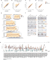
(A) Scatterplots of hydrolysis-based RNA-seq abundances (Log10 RPKM) of snRNAs and snoRNAs extended by 25 nt at the start and end coordinates. The x-axis shows RNA-seq abundances of wild-type L2 larvae (control wt rep1), the y-axis shows L2 homozygous mutants (E497K, Rexo5E497K, orange; PB, Rexo5PB, red) and wild-type L2 replicate 2 (control wt rep2, black). (B) Scatterplots of library-normalized read densities of 25-nt long windows at the 3′-termini of snoRNAs and snRNAs (3′ read density) for the same samples above. U8 snoRNA is marked (snoRNA:185/U8). (C,D) Same analysis as in (A,B) for tRNAs and tRNA 3′-termini. (E) Log10 RNA-seq coverage plots of selected snoRNAs for homozygous Rexo5PB (PB, red), Rexo5E497K (E497K, orange), and control L2 larvae (wt control rep2, black). Official gene name, nucleotide length (black) and type of transcription locus (intronic, independent) (red) are shown above each coverage plot, the length of the mature snoRNA (blue bar) below. The location of the mature snoRNA is shaded in orange; the 3′ precursor region upregulated in mutants is highlighted in yellow. Nucleotide position is given relative to transcript start site. U8 snoRNA (170 nt) extends beyond the predicted snoRNA:185 (54 nt) gene locus. The observed read coverage for U3 snoRNA (225 nt) extends the predicted length (168–173 nt). (F) Northern blot analysis of selected snoRNAs for Rexo5PB and Rexo5E497K homo- and heterozygous mutants and wild-type L2 larvae. Official gene names, lengths (black), and type (intronic, cistronic, independent) (red) are indicated below each blot. The same blot was stripped and re-probed for all probes. Loading control is 7SL RNA. (G) 3′ length analysis of snoRNA and snRNA precursors recorded by hydrolysis-based small RNA-seq, averaging six homozygous L2 mutants (three Rexo5PB and three Rexo5E497K pooled together) (red) and four wild-type L2 larvae (green). The x-axis shows snoRNA and snRNA gene identifiers, the y-axis the average number of nucleotides (nt) extending beyond the annotated transcript end (3′ length). Category 1: Selection of snoRNAs with the largest precursor 3′-processing changes accumulating in mutants. Category 2: Subgroup of highly abundant snoRNAs, which accumulate misprocessed precursors in mutants by density reads analysis in (B), but also have precursor coverage of >2 reads in wild-type controls. Category 3: subset of snRNAs. snRNAs show no altered 3′ end by length analysis and read density analysis.
Figure 4
Figure 4. Rexo5 mutants accumulate 3′-extended 5S rRNA
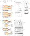
(A) Upper panel: Log10 RPKM read coverage plots of 5S rRNA in L2 homozygous mutant larvae (PB, Rexo5PB, red; E497K, Rexo5E497K, orange) and wild-type controls (black, both replicates) sequenced by hydrolysis-based RNA-seq. Lower panel: Log2 fold changes of read coverages (Log2 Diff) between mutants and wild-type control rep1 (E497K orange; PB red) and wild-type rep1 versus wild-type rep2 (black). Shown below is (1) length of the Flybase annotated 5S rRNA gene CR33395 (blue bar), (2) length of the observed 5S rRNA precursor (black) and (3) observed mature 5S rRNA by RNA-seq coverage (black). Mature 5S rRNA is shaded in orange, the 3′ trailer of the precursor in yellow. (B) Ethidium bromide stained 8% polyacrylamide/8M urea gel showing a higher molecular weight shift for 5S rRNA in L2 homozygous mutants and Tubulin-GAL4 UAS-shRNA-Rexo5 knockdowns. (C) Northern blot analysis for 5S, 5.8S rRNAs, 7SL RNA and U3 snoRNA in hetero- and homozygous Rexo5 mutants and wild type controls.
Figure 5
Figure 5. Rexo5 mutants accumulate 3′-ETS-containing 28S rRNA precursors
(A) Genomic map of rRNA precursors arranged in rDNA repeats. Location of the reference transcript M2017.1 used for alignments indicated with the dotted orange box. Upper panel: Hydrolysis-based RNA-seq read coverage (Log10 RPKM) of L2 homozygous mutants (PB, Rexo5PB red; E497K, Rexo5E497K, orange), and two replicate wild-type controls (gray). The location of mature 18S, 5.8S, 2S, and 28S rRNA is shown with the shaded orange areas. Lower panel: Log2-transformed coverage changes (Log2 Diff) between mutants and one wild-type control (same color code as in (A)) and coverage changes between two wild-type replicates (gray). (B) Same as in (A) for Illumina total RNA-seq (>200 nt read selection). (C) Canonical rRNA biogenesis pathway in D. melanogaster (Long and Dawid, 1980); cleavages sites are indicated with orange arrows. 28S rRNA hydrolytically fragments into 28Sa and 28Sb, which electrophoretically migrate close to 18S rRNA (Jordan, 1975). rRNA intermediates misprocessed in mutants are highlighted in bold red. (D) Northern blot analysis of rRNA precursors in homo- and heterozygous Rexo5E497K (E497K/E497K; E497K/+) and Rexo5PB (PB/PB; PB/+) and L2 wild-type larvae, probed for 5′-ETS, ITS1, mature 18S, mature 28Sb, 3′-ETS, and 7SL RNA as loading control. (E) Log10 RPKM read coverage plots along M2017.1 for in vivo Tubulin-GAL4 UAS-shRNA knockdowns (human ortholog in parentheses): Dbp73D (DDX51, red), nop5 (NOP58, blue), CG6937 (NIFK, green) and Rexo5 (LOC81691, orange), control shRNA knockdown of white (black), and wild-type L2 larvae (gray). Except for PAN2 and white, all knockdowns were lethal at L1 or L2. The 28S-3′-ETS transition is magnified in the box below. (F) Same as in (E) for 3′-5′ RNA exonucleases: Rexo5 (orange), Dis3 (red), Rrp6 (EXOSC10, blue), CG6833 (REXO4, yellow), genetic mutant CG12877e00300 (REXO1, green), PAN2 (gray), compared to control white (black). (G) Northern blot analysis probing for 3′-ETS, 5′-ETS, ITS1, 28S, 18S, and 7SL of shRNA knockdowns of nop5, RpS3, CG6937, Rexo5 and Dpb73D. All blots were stripped and re-probed for all probes.
Figure 6
Figure 6. Rexo5 homozygous mutants display increased nucleolar size and show nuclear accumulation of RpS2
(A) Confocal image of Rexo5 RNAi clones in the midgut of L3 larvae. Cells expressing UAS-shRNA Rexo5 express UAS-Red47 (red). The nucleolar protein Fibrillarin (Fib) is stained in green, DNA (Hoechst) in blue. RNAi UAS-Red47 cells are circled with a white dashed line. Genotype: hsFlp; Tubulin-GAL4/UAS-shRNA-Rexo5; hsFlp;Sco/Cyo;UAS#Red47a#1tub<+GFPPB/+) and (C-C”) homozygous (Rexo5PB/PB) L2 larvae. (B′, C) Gray scale images of 28S rRNA and (B”, C”) the 3′-ETS. (D–E) RNA-FISH probing against the 3′-ETS (red) and mature 18S rRNA (green) in (D-D”) heterozygous (Rexo5PB/+) and (E-E”) homozygous (Rexo5PB/PB) L2 larvae. (D′,E′) Gray scale images of 18S rRNA and (D”,E”) the 3′-ETS. (F–G) RNA-FISH probing against the 3′-ETS (red) in an RpS2-GFP (green) Rexo5PB genetic background. (F-F”) In vivo localization of RpS2-GFP (green) and the 3′-ETS (red) in (F) heterozygous (Rexo5PB/+) and (G) homozygous (Rexo5PB/PB) L2 mutant larvae. Nuclei are marked with Hoechst (blue). (F,G′) Gray scale images for RpS2-GFP and (F”,G”) the 3′-ETS.
Figure 7
Figure 7. Loss-of-function of Rexo5 causes mitotic and growth arrest in male and female gonads
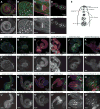
(A–D) Confocal imaging of transgenic GFP-Rexo5 under its native promoter; GFP-Rexo5 in green, DNA in blue (Hoechst), Phalloidin (A,C,D) or intracellular fusome marker 1B1 (B) are shown in red. (A–C) GFP-Rexo5 expression in adult testes (A,A′), at the testis apical tip (B,B′), in the cystic bulge (C,C). (D,D′) Rexo5 expression in adult ovarioles. (E) Schematic representation of the mitotic developmental program of Drosophila testis. Germ stem cells asymmetrically divide to produce one daughter stem cell and one gonialblast cell. The gonialblast migrates from the hub and undergoes four mitotic divisions. Stages of mutant and wild-type gonadal development at L2 are highlighted in red. (F,F) L2 wild-type male gonads stained for 1B1 (green) and DNA (blue). (F) 1B1 gray scale image. (G,G) Same staining as in (F,F′) for L2 Rexo5PB homozygous mutants. (H–M) Rexo5 mutants are lethal, but transgenic expression of pUASt-Rexo5 with Actin-GAL4 rescues somatic lethality to give adult flies that lack Rexo5 expression in germ cells (Brand and Perrimon, 1993). (H–I) 3-day-old adult wild-type and mutant testes. (H,H) Wild-type testes stained for actin (green, Phalloidin, PL), DNA (blue), and 1B1 (red). Mitotic cells (1B1) are restricted to the apical tip of the testis. (H) 1B1 gray scale image, (I,I′) Same as in (H,H) for Rexo5PB/E497K mutant testes. Mitotic cells are present throughout the entire testicular tube. (J,J′) 3-day old adult wild-type testis stained for Serine 10 phosphorylated histone H3 (PH3, red) marking meiotic nuclei. (J′) PH3 gray scale image. (K,K′) PH3 staining in mutant testes shows absence of meiotic nuclei. (L,L′) 3-day-old wild-type adult testis stained for phospho-H2av (red), Phalloidin (green), DNA (blue), (L′) gray scale image for H2Av. (M,M′) same as in (L,L′) for 3-day-old Actin-GAL4>Rexo5 Rexo5PB/E497K mutant testes. (N,N′) same as in (L,L′) for 14-day-old Actin-GAL4>Rexo5 rescued Rexo5E497K/Df(3L)BSC411 mutant testes. (O,O′) Wild-type ovarioles at 3 days, Actin-GAL4 UAS-Rexo5 Rexo5PBE497K mutant ovaries at 3 days (P,P′) and 14 days (Q,Q′), stained for Vasa (red), Phalloidin (green), DNA (blue).

References

    1. Allmang C, Kufel J, Chanfreau G, Mitchell P, Petfalski E, Tollervey D. Functions of the exosome in rRNA, snoRNA and snRNA synthesis. EMBO J. 1999;18:5399–5410. - PMC - PubMed
    1. Anderson MG, Perkins GL, Chittick P, Shrigley RJ, Johnson WA. drifter, a Drosophila POU-domain transcription factor, is required for correct differentiation and migration of tracheal cells and midline glia. Genes Dev. 1995;9:123–137. - PubMed
    1. Ansel KM, Pastor WA, Rath N, Lapan AD, Glasmacher E, Wolf C, Smith LC, Papadopoulou N, Lamperti ED, Tahiliani M, Ellwart JW, Shi Y, Kremmer E, Rao A, Heissmeyer V. Mouse Eri1 interacts with the ribosome and catalyzes 5.8S rRNA processing. Nat. Struct. Mol. Biol. 2008;15:523–530. - PMC - PubMed
    1. Berndt H, Harnisch C, Rammelt C, Stöhr N, Zirkel A, Dohm JC, Himmelbauer H, Tavanez J-P, Hüttelmaier S, Wahle E. Maturation of mammalian H/ACA box snoRNAs: PAPD5-dependent adenylation and PARN-dependent trimming. RNA. 2012;18:958–972. - PMC - PubMed
    1. Boulon S, Westman BJ, Hutten S, Boisvert F-M, Lamond AI. The Nucleolus under Stress. Mol. Cell. 2010;40:216–227. - PMC - PubMed

LinkOut - more resources