

Role of Condensing Particles in Polymer Confinement: A Model for Virus-Packed "Minichromosomes"
- PMID: 29045859
- PMCID: PMC5647577
- DOI: 10.1016/j.bpj.2017.08.035
Role of Condensing Particles in Polymer Confinement: A Model for Virus-Packed "Minichromosomes"
Abstract
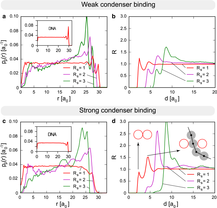
Confined mixtures of a polymer and nonspecifically binding particles (condensers) are studied as models for viruses containing double-stranded DNA (polymer) and condensing proteins (particles). We explore a model in which all interactions between the packed content (polymer and particles) and its confinement are purely repulsive, with only a short-range attraction between the condensers and polymer to simulate binding. In the range of physical parameters applicable to viruses, the model predicts reduction of pressure in the system effected by the condensers, despite the reduction in free volume. Condensers are found to be interspersed throughout the spherical confinement and only partially wrapped in the polymer, which acts as an effective medium for the condenser interactions. Crowding of the viral interior influences the DNA and protein organization, producing a picture inconsistent with a chromatin-like, beads-on-a-string structure. The model predicts an organization of the confined interior compatible with experimental data on unperturbed adenoviruses and polyomaviruses, at the same time providing insight into the role of condensing proteins in the viral infectious cycles of related viral families.
Copyright © 2017 Biophysical Society. Published by Elsevier Inc. All rights reserved.
Figures


Similar articles
-
Phage-like packing structures with mean field sequence dependence.J Comput Chem. 2017 Jun 5;38(15):1191-1197. doi: 10.1002/jcc.24727. Epub 2017 Mar 27. J Comput Chem. 2017. PMID: 28349552 Free PMC article.
-
Entropic attraction: Polymer compaction and expansion induced by nano-particles in confinement.J Chem Phys. 2015 May 7;142(17):174904. doi: 10.1063/1.4919650. J Chem Phys. 2015. PMID: 25956117
-
Trajectory Retracing of the Packaging and Ejection Processes of Coaxially Spooled DNA.J Chem Theory Comput. 2025 Jun 10;21(11):5736-5745. doi: 10.1021/acs.jctc.5c00137. Epub 2025 May 27. J Chem Theory Comput. 2025. PMID: 40432299
-
Analysis of X-ray diffraction from fibres of Pf1 Inovirus (filamentous bacteriophage) shows that the DNA in the virion is not highly ordered.J Mol Biol. 1998 Dec 18;284(5):1265-71. doi: 10.1006/jmbi.1998.2275. J Mol Biol. 1998. PMID: 9878347 Review.
-
Polymer models for the mechanisms of chromatin 3D folding: review and perspective.Phys Chem Chem Phys. 2020 Sep 23;22(36):20189-20201. doi: 10.1039/d0cp01877e. Phys Chem Chem Phys. 2020. PMID: 32966415 Review.
Cited by
-
Adenovirus core protein V reinforces the capsid and enhances genome release from disrupted particles.Sci Adv. 2023 Apr 7;9(14):eade9910. doi: 10.1126/sciadv.ade9910. Epub 2023 Apr 7. Sci Adv. 2023. PMID: 37027464 Free PMC article.
-
Maturation of Viruses.Subcell Biochem. 2024;105:503-531. doi: 10.1007/978-3-031-65187-8_14. Subcell Biochem. 2024. PMID: 39738956 Review.
-
pH stability and disassembly mechanism of wild-type simian virus 40.Soft Matter. 2020 Mar 21;16(11):2803-2814. doi: 10.1039/c9sm02436k. Epub 2020 Feb 27. Soft Matter. 2020. PMID: 32104873 Free PMC article.
-
Adenovirus Structure: What Is New?Int J Mol Sci. 2021 May 15;22(10):5240. doi: 10.3390/ijms22105240. Int J Mol Sci. 2021. PMID: 34063479 Free PMC article. Review.
-
Adenovirus major core protein condenses DNA in clusters and bundles, modulating genome release and capsid internal pressure.Nucleic Acids Res. 2019 Sep 26;47(17):9231-9242. doi: 10.1093/nar/gkz687. Nucleic Acids Res. 2019. PMID: 31396624 Free PMC article.
References
-
- Smith D.E., Tans S.J., Bustamante C. The bacteriophage straight ϕ29 portal motor can package DNA against a large internal force. Nature. 2001;413:748–752. - PubMed
-
- Šiber A., Božič A.L., Podgornik R. Energies and pressures in viruses: contribution of nonspecific electrostatic interactions. Phys. Chem. Chem. Phys. 2012;14:3746–3765. - PubMed
-
- Bloomfield V.A. DNA condensation by multivalent cations. Biopolymers. 1997;44:269–282. - PubMed
-
- Schiessel H. The physics of chromatin. J. Phys. Condens. Matter. 2003;15:R699–R774. - PubMed
MeSH terms
Substances
LinkOut - more resources
Full Text Sources
Other Literature Sources
