. 2017 Oct 17;113(8):1654-1663.
doi: 10.1016/j.bpj.2017.08.034.

Mechanics and Buckling of Biopolymeric Shells and Cell Nuclei

Affiliations

Mechanics and Buckling of Biopolymeric Shells and Cell Nuclei

Edward J Banigan et al. Biophys J. .

Abstract

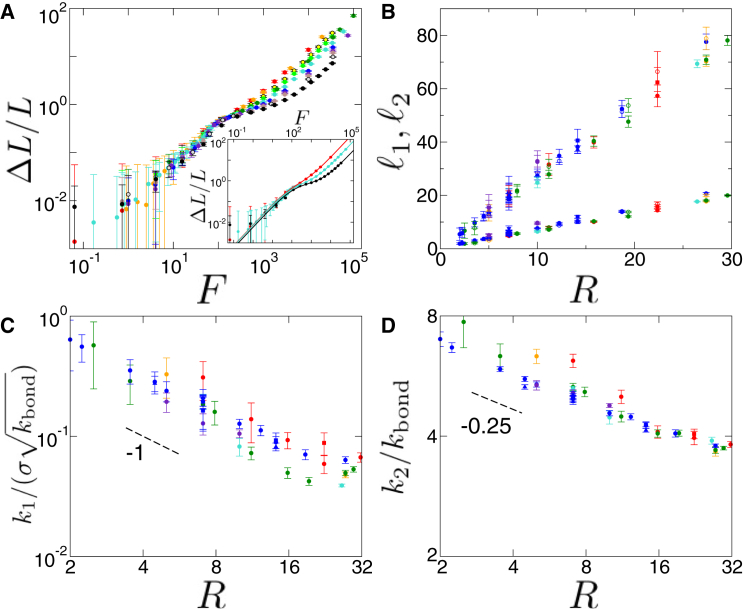
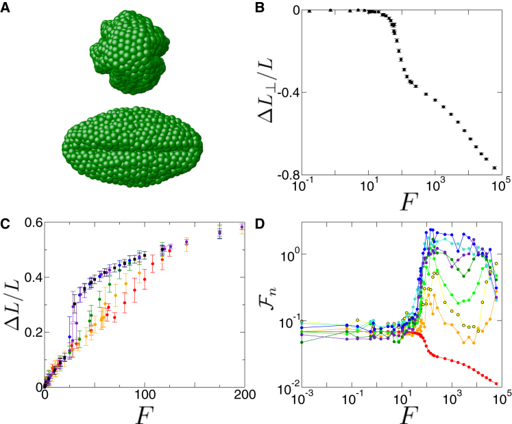
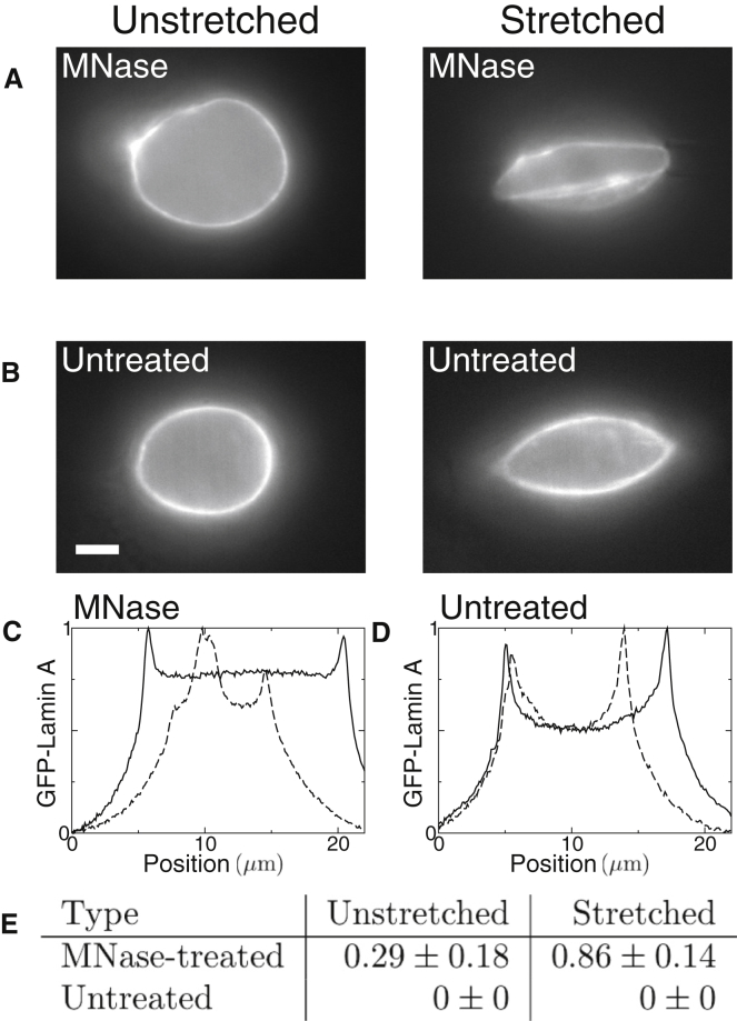
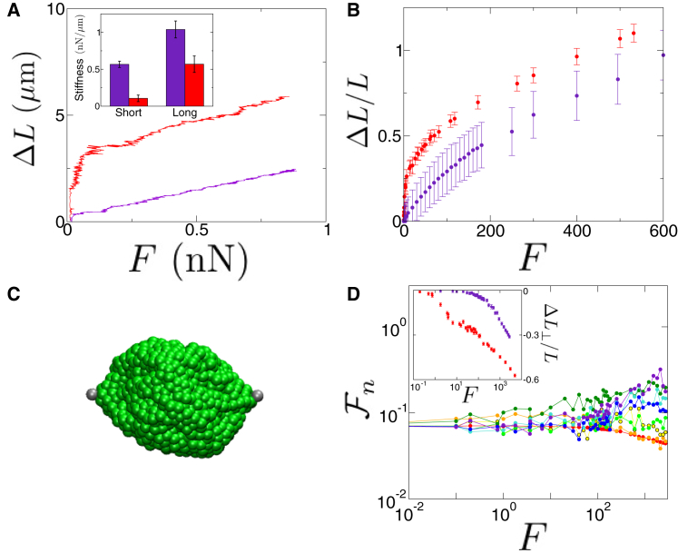
We study a Brownian dynamics simulation model of a biopolymeric shell deformed by axial forces exerted at opposing poles. The model exhibits two distinct, linear force-extension regimes, with the response to small tensions governed by linear elasticity and the response to large tensions governed by an effective spring constant that scales with radius as R-0.25. When extended beyond the initial linear elastic regime, the shell undergoes a hysteretic, temperature-dependent buckling transition. We experimentally observe this buckling transition by stretching and imaging the lamina of isolated cell nuclei. Furthermore, the interior contents of the shell can alter mechanical response and buckling, which we show by simulating a model for the nucleus that quantitatively agrees with our micromanipulation experiments stretching individual nuclei.


Figures

Figure 1
Figure 1
Tension-strain relation for polymeric shells. (A) Shell stretched by tension F exhibits two-regime response, with linear response at small tensions and stiffer linear response to large tensions. Tension-strain curves do not scale simply with N (σN/R2=20 with N=80, red; 100, orange; 250, yellow; 400, light green; 500, dark green; 1000, turquoise; 2000, blue; 3000, purple, 4000, brown; 7000, gray; 15,000, black). Inset: Examples of fitting by Eq. 3. (B) Crossover lengths, 1 and 2, scale linearly with shell radius, R. Colors, shapes, and filling of symbols indicate different σ, k, and kBT, respectively (σ=2, red; 4, orange; 8, dark green; 10, turquoise; 20, blue; 40, purplek=10, downward pointing triangle; 25, square; 50, diamond; 100, circle; 200, triangle up; 400, triangle left. kBT=103, filled; 1, open). (C) Small extension spring constant scales as k1/(σkbond)R1. (D) Large extension spring constant scales as k2/kbondR0.25. Symbols for (C) and (D) are as in (B). Lengths are in units of a, tensions are in simulation force units of f0, spring constants are in units of f0/a, and temperatures are in units of f0a. Measurements are from at least 11 simulations per data point. To see this figure in color, go online.
Figure 2
Figure 2
Buckling of stretched polymeric shells. (A) Two views of stretched shell with multiple buckles perpendicular to tension axis extending longitudinally across the shell. (B) Transverse strain, ΔL/L, versus tension, F, drops sharply at the buckling transition. (C) Tension-strain relation exhibits a signature of the buckling transition, with a jump in strain that increases with increasing temperature (kBT=0 (no Langevin noise, see Materials and Methods), red; 103, orange; 102, green; 101, blue; 1, purple). Black squares show hysteretic behavior of tension-strain relation for shells evolved at kBT=103 after a transient of kBT=1. (D) Normalized Fourier modes, Fn(F) (Eqs. 8 and 9), indicate buckling by increases in modes with n2 at tensions corresponding to the jump in the tension-strain curve (n=0, red; 2, orange; 3, yellow; 4, light green; 5, dark green; 6, turquoise; 7, blue; 8, purple). To see this figure in color, go online.
Figure 3
Figure 3
Stretched laminas, but not the nuclei containing chromatin, exhibit axial buckles. (A) Representative images of an unstretched (left) and stretched (right) lamina, obtained by treating HeLa nuclei with MNase, which digests chromatin. Stretched image shows longitudinal buckles. (B) Representative images of nuclei with intact chromatin interior, which do not exhibit buckling when stretched (right; compare to left, unstretched). Scale bars represent 5μm. Fluorescence signal is GFP-lamin A. (C) Line scans along the central axis perpendicular to the tension axis showing the GFP-lamin A signal for an MNase-treated nucleus when unstretched (solid line) and stretched (dashed line), corresponding to the images in (A). GFP-lamin A intensity is plotted in arbitrary units. (D) Line scans along the central axis perpendicular to the tension axis showing the GFP-lamin A signal for an untreated nucleus when unstretched (solid line) and stretched (dashed line), corresponding to the images in (B). (E) Table listing average number of excess peaks (between boundary peaks) per line scan. Seven nuclei were imaged for each case. Error is given by SE.
Figure 4
Figure 4
Shells filled with a tethered cross-linked polymer, modeling a cell nucleus, do not axially buckle. (A) Force-extension curves for MEF-V−/− nuclei in experiments in which nuclei were treated with MNase (red) and without (purple) MNase treatment. Inset: Bar graph showing effective spring constants for MEF-V−/− nuclei in short and long extension regimes with (red) and without (purple) MNase treatment. Measurements were performed for 18 untreated and five MNase-treated nuclei. Error bars are mean ± SE. Statistical significance of p<0.05 was established by t-test. (B) Tension-strain relation shows that the tethered cross-linked polymer interior strengthens initial force response (purple, as compared to empty shell, red). Simulation units are a=0.7μm and f0=5.9pN. (C) Although there are localized buckles in the stretched shell with a tethered polymer interior, buckles do not span the entire structure. (D) Normalized Fourier modes show weaker signature of buckling, compared to those of Fig. 2D (n=0, red; 2, orange; 3, yellow; 4, light green; 5, dark green; 6, turquoise; 7, blue; 8, purple). Inset: Transverse strain for shells enclosing a tethered polymer (purple) and empty shells (red). z=4(z4.5) in (B)–(D). To see this figure in color, go online.

References

    1. Lavrentovich M.O., Horsley E.M., Kamien R.D. First-order patterning transitions on a sphere as a route to cell morphology. Proc. Natl. Acad. Sci. USA. 2016;113:5189–5194. - PMC - PubMed
    1. Lidmar J., Mirny L., Nelson D.R. Virus shapes and buckling transitions in spherical shells. Phys. Rev. E Stat. Nonlin. Soft Matter Phys. 2003;68:051910. - PubMed
    1. Šiber A. Buckling transition in icosahedral shells subjected to volume conservation constraint and pressure: relations to virus maturation. Phys. Rev. E Stat. Nonlin. Soft Matter Phys. 2006;73:061915. - PubMed
    1. Arnoldi M., Fritz M., Boulbitch A. Bacterial turgor pressure can be measured by atomic force microscopy. Phys. Rev. E Stat. Phys. Plasmas Fluids Relat. Interdiscip. Topics. 2000;62(1 Pt B):1034–1044. - PubMed
    1. Crowther R.A., Finch J.T., Pearse B.M.F. On the structure of coated vesicles. J. Mol. Biol. 1976;103:785–798. - PubMed

LinkOut - more resources