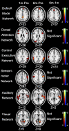
Re-Establishing Brain Networks in Patients with ESRD after Successful Kidney Transplantation
- PMID: 29046290
- PMCID: PMC5753300
- DOI: 10.2215/CJN.00420117
Re-Establishing Brain Networks in Patients with ESRD after Successful Kidney Transplantation
Expression of concern in
-
Expression of Concern: Re-Establishing Brain Networks in Patients with ESRD after Successful Kidney Transplantation.Clin J Am Soc Nephrol. 2019 Jul 5;14(7):1072. doi: 10.2215/CJN.04190419. Epub 2019 Apr 24. Clin J Am Soc Nephrol. 2019. PMID: 31018933 Free PMC article. No abstract available.
Abstract
Background and objectives: Cognition in ESRD may be improved by kidney transplantation, but mechanisms are unclear. We explored patterns of resting-state networks with resting-state functional magnetic resonance imaging among patients with ESRD before and after kidney transplantation.
Design, setting, participants, & measurements: Thirty-seven patients with ESRD scheduled for kidney transplantation and 22 age-, sex-, and education-matched healthy subjects underwent resting-state functional magnetic resonance imaging. Patients were imaged before and 1 and 6 months after kidney transplantation. Functional connectivity of seven resting-state subnetworks was evaluated: default mode network, dorsal attention network, central executive network, self-referential network, sensorimotor network, visual network, and auditory network. Mixed effects models tested associations of ESRD, kidney transplantation, and neuropsychological measurements with functional connectivity.
Results: Compared with controls, pretransplant patients showed abnormal functional connectivity in six subnetworks. Compared with pretransplant patients, increased functional connectivity was observed in the default mode network, the dorsal attention network, the central executive network, the sensorimotor network, the auditory network, and the visual network 1 and 6 months after kidney transplantation (P=0.01). Six months after kidney transplantation, no significant difference in functional connectivity was observed for the dorsal attention network, the central executive network, the auditory network, or the visual network between patients and controls. Default mode network and sensorimotor network remained significantly different from those in controls when assessed 6 months after kidney transplantation. A relationship between functional connectivity and neuropsychological measurements was found in specific brain regions of some brain networks.
Conclusions: The recovery patterns of resting-state subnetworks vary after kidney transplantation. The dorsal attention network, the central executive network, the auditory network, and the visual network recovered to normal levels, whereas the default mode network and the sensorimotor network did not recover completely 6 months after kidney transplantation. Neural resting-state functional connectivity was lower among patients with ESRD compared with control subjects, but it significantly improved with kidney transplantation. Resting-state subnetworks exhibited variable recovery, in some cases to levels that were no longer significantly different from those of normal controls.
Keywords: Attention; Brain; Cognition; Healthy Volunteers; Kidney Failure, Chronic; Magnetic Resonance Imaging; end-stage renal disease; kidney transplantation.
Copyright © 2018 by the American Society of Nephrology.
Figures


Comment in
-
Trust Patient Insights at Both the Individual and National Level.Clin J Am Soc Nephrol. 2018 Jan 6;13(1):1-2. doi: 10.2215/CJN.13211117. Epub 2017 Dec 21. Clin J Am Soc Nephrol. 2018. PMID: 29269565 Free PMC article. No abstract available.
References
-
- Bugnicourt JM, Godefroy O, Chillon JM, Choukroun G, Massy ZA: Cognitive disorders and dementia in CKD: The neglected kidney-brain axis. J Am Soc Nephrol 24: 353–363, 2013 - PubMed
-
- Hermann DM, Kribben A, Bruck H: Cognitive impairment in chronic kidney disease: Clinical findings, risk factors and consequences for patient care. J Neural Transm (Vienna) 121: 627–632, 2014 - PubMed
-
- Kurella M, Chertow GM, Luan J, Yaffe K: Cognitive impairment in chronic kidney disease. J Am Geriatr Soc 52: 1863–1869, 2004 - PubMed
-
- Fazekas G, Fazekas F, Schmidt R, Kapeller P, Offenbacher H, Krejs GJ: Brain MRI findings and cognitive impairment in patients undergoing chronic hemodialysis treatment. J Neurol Sci 134: 83–88, 1995 - PubMed
-
- Murray AM, Tupper DE, Knopman DS, Gilbertson DT, Pederson SL, Li S, Smith GE, Hochhalter AK, Collins AJ, Kane RL: Cognitive impairment in hemodialysis patients is common. Neurology 67: 216–223, 2006 - PubMed
Publication types
MeSH terms
LinkOut - more resources
Full Text Sources
Other Literature Sources
Medical

