

Bone mineral density during pregnancy in women participating in a randomized controlled trial of vitamin D supplementation
- PMID: 29046301
- PMCID: PMC5698834
- DOI: 10.3945/ajcn.116.140459
Bone mineral density during pregnancy in women participating in a randomized controlled trial of vitamin D supplementation
Abstract
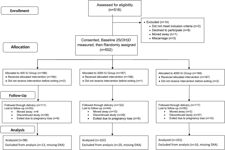
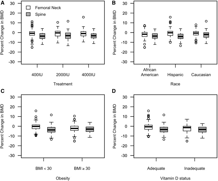
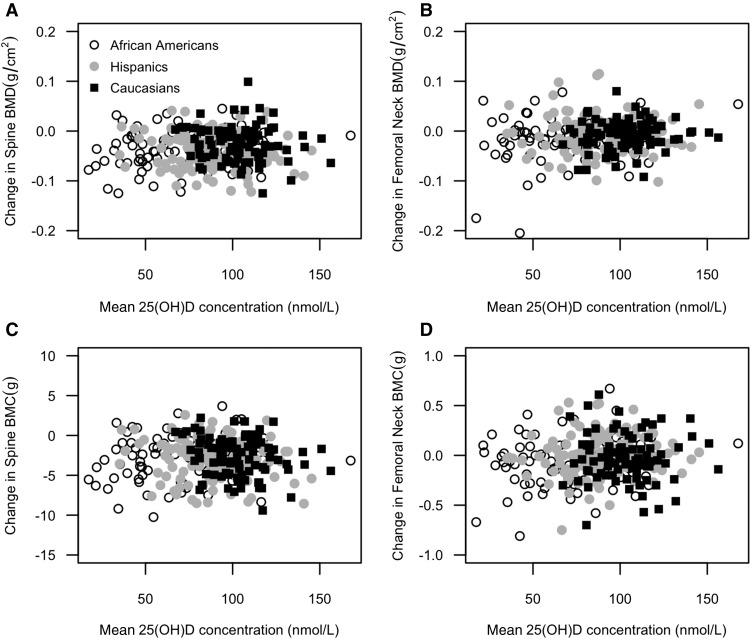
Background: Little is known about bone mineral density (BMD) during pregnancy. Advances in technology with lower radiation emissions by dual-energy X-ray absorptiometry instruments now permit the safe measurement of BMD during pregnancy.Objective: We evaluated maternal BMD during pregnancy as a function of vitamin D status in women of diverse racial/ethnic backgrounds.Design: A total of 301 women who underwent BMD measurements at 12-20 wk of gestation and again at 0-14 wk postpartum were included in this analysis. Women were a subset of subjects who were recruited for a randomized, controlled, double-blind trial of vitamin D supplementation in pregnancy (400, 2000, or 4000 IU/d).Results: Treatment had no significant effect on changes in BMD that occurred between 12-20 wk of gestation and 0-14 wk postpartum. Similarly, changes in spine and femoral neck bone mineral contents (BMCs) were not significantly different in the treatment groups. In addition, vitamin D inadequacy (serum 25-hydroxyvitamin D concentration, averaged across pregnancy, <50 nmol/L) was not associated with changes in BMD or BMC. There were significant racial/ethnic differences in spine BMD. African Americans lost more spine BMD than did Caucasians (-0.04 ± 0.04 compared with -0.02 ± 0.04 g/cm2; P = 0.033). In addition, baseline obesity was associated with a greater loss of femoral neck BMD. The means ± SDs of femoral neck BMD loss were -0.02 ± 0.05 and 0.0 ± 0.03 g/cm2 for groups with baseline body mass index (BMI; in kg/m2) ≥30 and <30, respectively.Conclusion: These findings do not support a dose effect of vitamin D supplementation on bone health and suggest that race/ethnicity and BMI play an important role in pregnancy bone health. This trial was registered at clinicaltrials.gov as NCT00292591.
Keywords: bone mineral content; bone mineral density; cholecalciferol; pregnancy; vitamin D.
© 2017 American Society for Nutrition.
Figures
Similar articles
-
Factors Affecting Postpartum Bone Mineral Density in a Clinical Trial of Vitamin D Supplementation.J Womens Health (Larchmt). 2024 Jul;33(7):887-900. doi: 10.1089/jwh.2022.0525. Epub 2024 Jun 10. J Womens Health (Larchmt). 2024. PMID: 38853682 Clinical Trial.
-
Effect of calcium plus vitamin D supplementation during pregnancy in Brazilian adolescent mothers: a randomized, placebo-controlled trial.Am J Clin Nutr. 2013 Jul;98(1):82-91. doi: 10.3945/ajcn.112.056275. Epub 2013 May 29. Am J Clin Nutr. 2013. PMID: 23719547 Clinical Trial.
-
Calcium Plus Vitamin D Supplementation During the Third Trimester of Pregnancy in Adolescents Accustomed to Low Calcium Diets Does Not Affect Infant Bone Mass at Early Lactation in a Randomized Controlled Trial.J Nutr. 2015 Jul;145(7):1515-23. doi: 10.3945/jn.114.208140. Epub 2015 May 27. J Nutr. 2015. PMID: 26019245 Clinical Trial.
-
The effect of pregnancy vitamin D supplementation on offspring bone mineral density in childhood: a systematic review and meta-analysis.Osteoporos Int. 2023 Jul;34(7):1269-1279. doi: 10.1007/s00198-023-06751-5. Epub 2023 Apr 27. Osteoporos Int. 2023. PMID: 37103591
-
Vitamin D supplementation for improving bone density in vitamin D-deficient children and adolescents: systematic review and individual participant data meta-analysis of randomized controlled trials.Am J Clin Nutr. 2023 Sep;118(3):498-506. doi: 10.1016/j.ajcnut.2023.05.028. Epub 2023 Aug 8. Am J Clin Nutr. 2023. PMID: 37661104
Cited by
-
Effect of prenatal calcium supplementation on bone during pregnancy and 1 y postpartum.Am J Clin Nutr. 2019 Jan 1;109(1):197-206. doi: 10.1093/ajcn/nqy233. Am J Clin Nutr. 2019. PMID: 30649176 Free PMC article. Clinical Trial.
-
25-Hydroxyvitamin D profiles and maternal bone mass during pregnancy and lactation in Japanese women.J Bone Miner Metab. 2020 Jan;38(1):99-108. doi: 10.1007/s00774-019-01032-w. Epub 2019 Aug 20. J Bone Miner Metab. 2020. PMID: 31432265
-
Vitamin D supplementation for women during pregnancy.Cochrane Database Syst Rev. 2019 Jul 26;7(7):CD008873. doi: 10.1002/14651858.CD008873.pub4. Cochrane Database Syst Rev. 2019. Update in: Cochrane Database Syst Rev. 2024 Jul 30;7:CD008873. doi: 10.1002/14651858.CD008873.pub5. PMID: 31348529 Free PMC article. Updated.
-
The association of homocysteine, folate, vitamin B12, and vitamin B6 with fracture incidence in older adults: a systematic review and meta-analysis.Ann Transl Med. 2021 Jul;9(14):1143. doi: 10.21037/atm-21-2514. Ann Transl Med. 2021. PMID: 34430584 Free PMC article.
-
Does parity and duration of lactation have any effect on the bone mineral density of the femur and lumbar spine in Indian women? A cross-sectional study from the Northeast region of India.J Family Med Prim Care. 2021 Aug;10(8):2886-2892. doi: 10.4103/jfmpc.jfmpc_2349_20. Epub 2021 Aug 27. J Family Med Prim Care. 2021. PMID: 34660421 Free PMC article.
References
-
- Drinkwater BL, Chesnut CH III. Bone density changes during pregnancy and lactation in active women: a longitudinal study. Bone Miner 1991;14:153–60. - PubMed
-
- Naylor KE, Iqbal P, Fledelius C, Fraser RB, Eastell R. The effect of pregnancy on bone density and bone turnover. J Bone Miner Res 2000;15:129–37. - PubMed
-
- Kaur M, Pearson D, Godber I, Lawson N, Baker P, Hosking D. Longitudinal changes in bone mineral density during normal pregnancy. Bone 2003;32:449–54. - PubMed
-
- Dahlman I, Gerdhem P, Bergstrom I. Vitamin D status and bone health in immigrant versus Swedish women during pregnancy and the post-partum period. J Musculoskelet Neuronal Interact 2013;13:464–9. - PubMed
Publication types
MeSH terms
Substances
Associated data
LinkOut - more resources
Full Text Sources
Other Literature Sources
Medical
