

Respiratory dysfunction following neonatal sustained hypoxia exposure during a critical window of brain stem extracellular matrix formation
- PMID: 29046314
- PMCID: PMC5867672
- DOI: 10.1152/ajpregu.00199.2017
Respiratory dysfunction following neonatal sustained hypoxia exposure during a critical window of brain stem extracellular matrix formation
Abstract
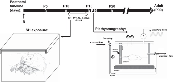
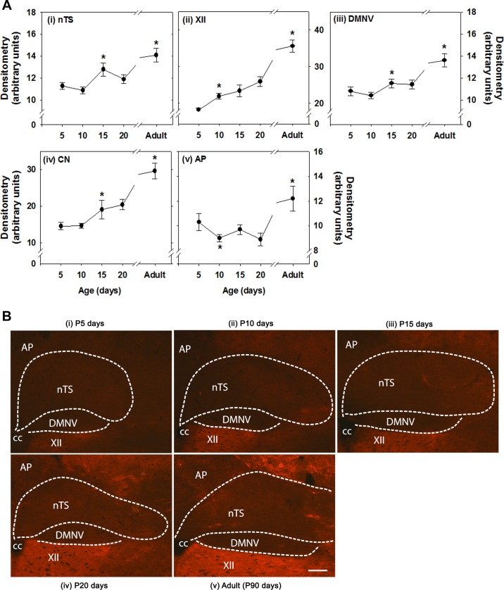
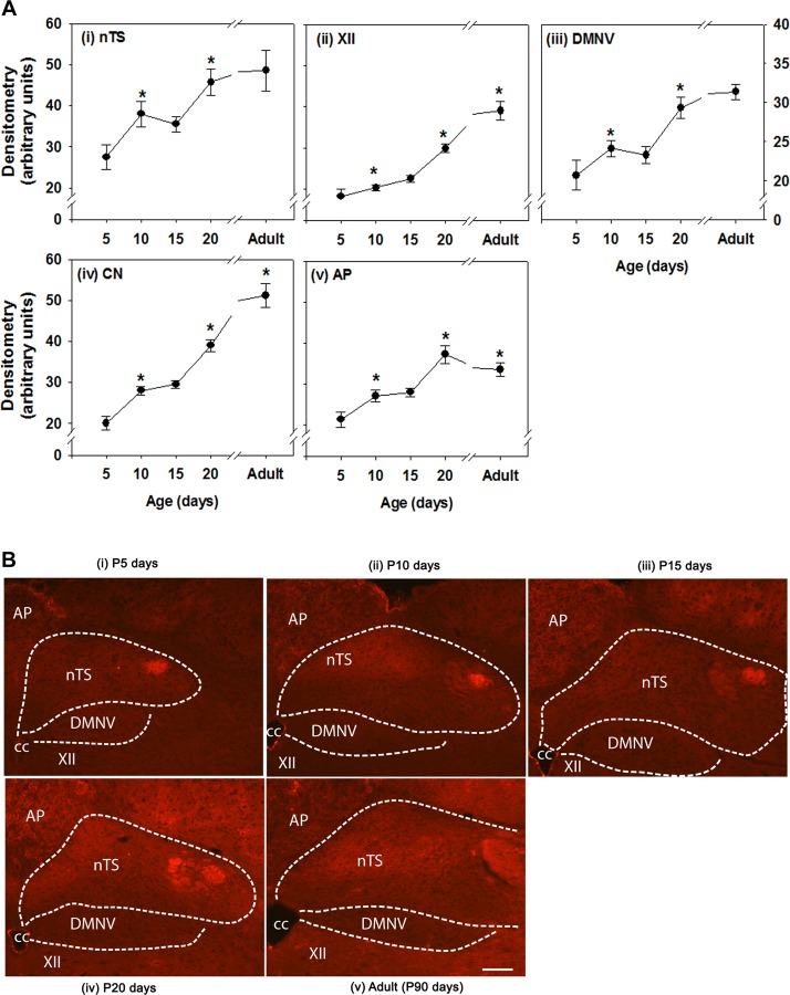
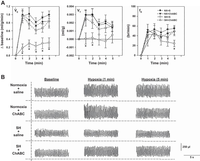
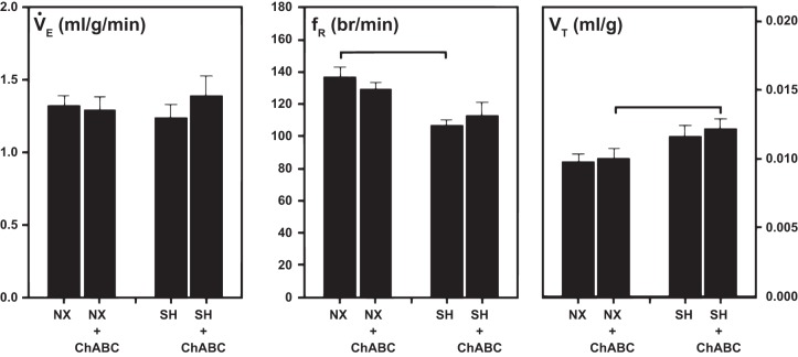
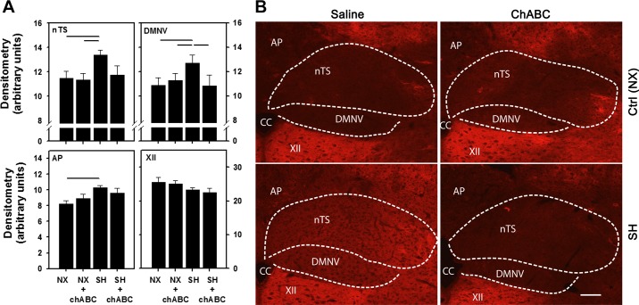
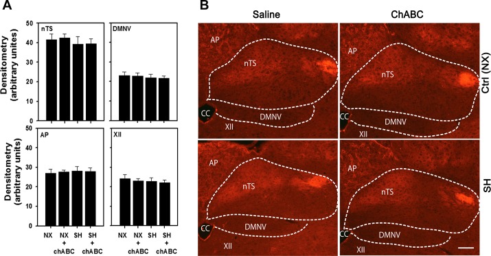
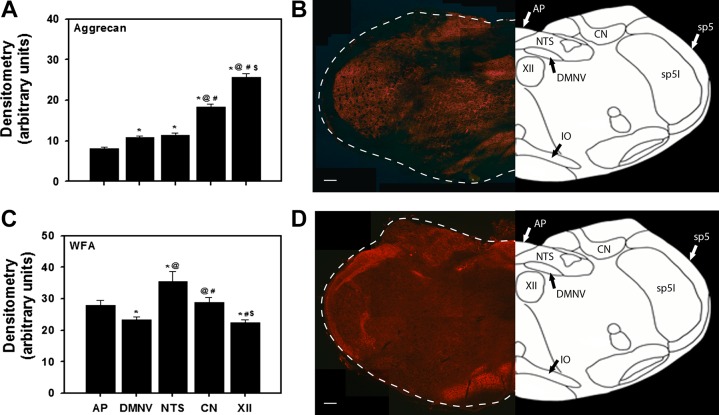
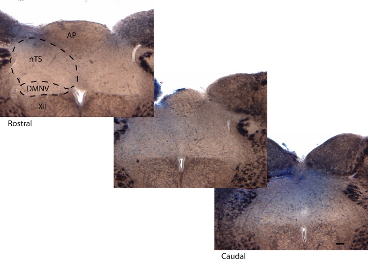
The extracellular matrix (ECM) modulates brain maturation and plays a major role in regulating neuronal plasticity during critical periods of development. We examined 1) whether there is a critical postnatal period of ECM expression in brain stem cardiorespiratory control regions and 2) whether the attenuated hypoxic ventilatory response (HVR) following neonatal sustained (5 days) hypoxia [SH (11% O2, 24 h/day)] exposure is associated with altered ECM formation. The nucleus tractus solitarius (nTS), dorsal motor nucleus of the vagus, hypoglossal motor nucleus, cuneate nucleus, and area postrema were immunofluorescently processed for aggrecan and Wisteria floribunda agglutinin (WFA), a key proteoglycan of the ECM and the perineuronal net. From postnatal day ( P) 5 ( P5), aggrecan and WFA expression increased postnatally in all regions. We observed an abrupt increase in aggrecan expression in the nTS, a region that integrates and receives afferent inputs from the carotid body, between P10 and P15 followed by a distinct and transient plateau between P15 and P20. WFA expression in the nTS exhibited an analogous transient plateau, but it occurred earlier (between P10 and P15). SH between P11 and P15 attenuated the HVR (assessed at P16) and increased aggrecan (but not WFA) expression in the nTS, dorsal motor nucleus of the vagus, and area postrema. An intracisternal microinjection of chondroitinase ABC, an enzyme that digests chondroitin sulfate proteoglycans, rescued the HVR and the increased aggrecan expression. These data indicate that important stages of ECM formation take place in key brain stem respiratory neural control regions and appear to be associated with a heightened vulnerability to hypoxia.
Keywords: brain stem extracellular matrix; breathing; development; hypoxia.
Figures
Similar articles
-
Experience-dependent development of perineuronal nets and chondroitin sulfate proteoglycan receptors in mouse visual cortex.Matrix Biol. 2013 Aug 8;32(6):352-63. doi: 10.1016/j.matbio.2013.04.001. Epub 2013 Apr 15. Matrix Biol. 2013. PMID: 23597636
-
Microglia modulate brainstem serotonergic expression following neonatal sustained hypoxia exposure: implications for sudden infant death syndrome.J Physiol. 2016 Jun 1;594(11):3079-94. doi: 10.1113/JP271845. Epub 2016 Feb 21. J Physiol. 2016. PMID: 26659585 Free PMC article.
-
Sensory experience-dependent formation of perineuronal nets and expression of Cat-315 immunoreactive components in the mouse somatosensory cortex.Neuroscience. 2017 Jul 4;355:161-174. doi: 10.1016/j.neuroscience.2017.04.041. Epub 2017 May 8. Neuroscience. 2017. PMID: 28495333
-
AMPK breathing and oxygen supply.Respir Physiol Neurobiol. 2019 Jul;265:112-120. doi: 10.1016/j.resp.2018.08.011. Epub 2018 Sep 19. Respir Physiol Neurobiol. 2019. PMID: 30243821 Review.
-
Neural Processing without O2 and Glucose Delivery: Lessons from the Pond to the Clinic.Physiology (Bethesda). 2024 Nov 1;39(6):0. doi: 10.1152/physiol.00030.2023. Epub 2024 Apr 16. Physiology (Bethesda). 2024. PMID: 38624246 Review.
Cited by
-
Disruption of rat deep cerebellar perineuronal net alters eyeblink conditioning and neuronal electrophysiology.Neurobiol Learn Mem. 2021 Jan;177:107358. doi: 10.1016/j.nlm.2020.107358. Epub 2020 Dec 4. Neurobiol Learn Mem. 2021. PMID: 33285318 Free PMC article.
-
Ventilatory and carotid body responses to acute hypoxia in rats exposed to chronic hypoxia during the first and second postnatal weeks.Respir Physiol Neurobiol. 2020 Apr;275:103400. doi: 10.1016/j.resp.2020.103400. Epub 2020 Jan 30. Respir Physiol Neurobiol. 2020. PMID: 32006667 Free PMC article.
-
Rodent models of respiratory control and respiratory system development-Clinical significance.Respir Physiol Neurobiol. 2019 Oct;268:103249. doi: 10.1016/j.resp.2019.06.006. Epub 2019 Jul 14. Respir Physiol Neurobiol. 2019. PMID: 31315068 Free PMC article. Review.
-
Mechanisms underlying a critical period of respiratory development in the rat.Respir Physiol Neurobiol. 2019 Jun;264:40-50. doi: 10.1016/j.resp.2019.04.006. Epub 2019 Apr 15. Respir Physiol Neurobiol. 2019. PMID: 30999061 Free PMC article. Review.
-
Mechanistic actions of oxygen and methylxanthines on respiratory neural control and for the treatment of neonatal apnea.Respir Physiol Neurobiol. 2020 Feb;273:103318. doi: 10.1016/j.resp.2019.103318. Epub 2019 Oct 15. Respir Physiol Neurobiol. 2020. PMID: 31626973 Free PMC article. Review.
References
-
- Brückner G, Brauer K, Härtig W, Wolff JR, Rickmann MJ, Derouiche A, Delpech B, Girard N, Oertel WH, Reichenbach A. Perineuronal nets provide a polyanionic, glia-associated form of microenvironment around certain neurons in many parts of the rat brain. Glia 8: 183–200, 1993. doi:10.1002/glia.440080306. - DOI - PubMed
Publication types
MeSH terms
Substances
LinkOut - more resources
Full Text Sources
Other Literature Sources
Medical
Miscellaneous