Comparative Study
. 2017 Dec 15;292(50):20449-20460.
doi: 10.1074/jbc.M117.811182. Epub 2017 Oct 18.

Evidence for a conserved inhibitory binding mode between the membrane fusion assembly factors Munc18 and syntaxin in animals

Affiliations
Comparative Study

Evidence for a conserved inhibitory binding mode between the membrane fusion assembly factors Munc18 and syntaxin in animals

Czuee Morey et al. J Biol Chem. .

Abstract

The membrane fusion necessary for vesicle trafficking is driven by the assembly of heterologous SNARE proteins orchestrated by the binding of Sec1/Munc18 (SM) proteins to specific syntaxin SNARE proteins. However, the precise mode of interaction between SM proteins and SNAREs is debated, as contrasting binding modes have been found for different members of the SM protein family, including the three vertebrate Munc18 isoforms. While different binding modes could be necessary, given their roles in different secretory processes in different tissues, the structural similarity of the three isoforms makes this divergence perplexing. Although the neuronal isoform Munc18a is well-established to bind tightly to both the closed conformation and the N-peptide of syntaxin 1a, thereby inhibiting SNARE complex formation, Munc18b and -c, which have a more widespread distribution, are reported to mainly interact with the N-peptide of their partnering syntaxins and are thought to instead promote SNARE complex formation. We have reinvestigated the interaction between Munc18c and syntaxin 4 (Syx4). Using isothermal titration calorimetry, we found that Munc18c, like Munc18a, binds to both the closed conformation and the N-peptide of Syx4. Furthermore, using a novel kinetic approach, we found that Munc18c, like Munc18a, slows down SNARE complex formation through high-affinity binding to syntaxin. This strongly suggests that secretory Munc18s in general control the accessibility of the bound syntaxin, probably preparing it for SNARE complex assembly.

Keywords: Munc18; SM protein; SNARE proteins; glucose transporter type 4 (GLUT4); insulin; membrane transport; secretion; syntaxin.


Conflict of interest statement

The authors declare that they have no conflicts of interest with the contents of this article

Figures

Figure 1.
Figure 1.
Schematic outline of an evolutionary tree of the secretory SM protein Munc18 in animals. The tree shows that genome duplications in vertebrates resulted in three distinct Munc18 paralogs (Munc18a, Munc18b, and Munc18c) that are separated from other animal Munc18s. An additional duplication of Munc18a occurred in teleosts. Note that many other animals have only a single Munc18 gene. The tree was constructed from 551 sequences derived from 253 animal species (including 181 vertebrates) and three unicellular holozoans (supplemental Table S1). Statistical support values (IQ-TREE support and RAxML support) are given at selected inner edges. The original tree file is available in Nexus format as supplemental Fig. S6. The following crystal structures are shown at the corresponding positions in the tree: rat Munc18a-Syx1a complex (PDB code 3C98) (10), human Munc18b (PDB code 4CCA) (47), Munc18c-Syx4 N-peptide complex (PDB code 3PUK) (49), squid Sec1 (PDB code 1EPU) (97), and choanoflagellate Munc18-Syx1 complex (PDB code 2XHE) (11). Note that only the structures of rat Munc18a and that of the choanoflagellate Monosiga brevicollis have been determined in complexes with the closed conformation of syntaxin and the N-peptide. The four-helix bundle of the closed syntaxin is accommodated in the cavity between domains 1 and 3a.
Figure 2.
Figure 2.
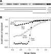
The N-peptide and the remainder of Syx4 contribute to binding to Munc18c. A, schematic drawing of the domain structure of Syx4 and fragments used in this experiment. The short N-peptide (NP) motif, the three Habc helices, the SNARE motif, and the transmembrane region (TMR) are indicated. B, calorimetric titrations of different Syx4 variants into Munc18c. The integrated areas normalized to the amount of Syx4 (kcal/mol) versus the molar ratio of Syx4 to Munc18c are shown. The solid lines represent the best fit to the data for a single binding site model using nonlinear least squares fitting. The affinity measured for binding of the soluble portion of Syx4 to Munc18c via ITC is about 12 nm. It also corroborates, as remarked earlier (58), that binding between Munc18c and Syx4 is somewhat weaker than between the neuronal pair Munc18a and Syx1a (Kd ≈ 1 nm (10)). Of note, the affinity determined by us is higher than that reported previously via ITC (58) (Kd ≈ 95 nm), whereas an affinity of 32 nm had been determined by surface plasmon resonance (66). Note that the corresponding data are given in Table 1 and that all ITC experiments are shown in supplemental Fig. S2.
Figure 3.
Figure 3.
Munc18c inhibits SNARE complex formation of bound Syx4. Ternary SNARE complex formation was followed by an increase in fluorescence anisotropy of 40 nm fluorescent Syb(1–96)61OG upon mixing with 1 μm Syx4(1–270) and 1.5 μm SNAP-25. In the presence of 1.5 μm Munc18c, ternary SNARE complex formation was slowed down.
Figure 4.
Figure 4.
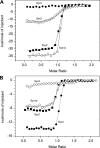
Munc18a and Munc18c can interact with different syntaxins. Shown is calorimetric titration of different secretory syntaxins into Munc18a (A) and Munc18c (B). Note that the titration of Syx4 into Munc18c is also shown in Fig. 2. Munc18a can interact tightly with Syx1a and Syx2 but binds only with moderate affinity to Syx3. Munc18c can interact tightly with Syx1a, Syx2, and Syx4. Munc18a does not interact with Syx4, whereas Munc18c does not interact with Syx3.
Figure 5.
Figure 5.
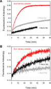
Munc18a and Munc18c inhibit the SNARE complex formation of bound Syx2 and Syx1a. Ternary SNARE complex formation was followed by an increase in the fluorescence anisotropy of 40 nm fluorescent Syb(1–96)61OG upon mixing with SNAP25 and the soluble portion of Syx2 (Syx2(1–262); A) or Syx1a (Syx1a(1–262); B). For the formation of the Syx1a SNARE complex, 0.5 μm Syx1a and 0.75 μm SNAP-25 were mixed. In the presence of 0.75 μm Munc18a or Munc18c, SNARE complex formation was slowed down. For the assembly of the Syx2 SNARE complex, 2 μm Syx2 and 3 μm SNAP-25 were mixed. In the presence of 3 μm Munc18a or Munc18c in the reaction mix, SNARE complex formation was slowed down.
Figure 6.
Figure 6.
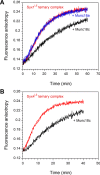
Munc18c inhibits the SNARE complex formation of SyxLE variants. Ternary SNARE complex formation was followed by an increase in the fluorescence anisotropy of 40 nm fluorescent Syb(1–96)61OG upon mixing with SNAP25 and the LE mutants of Syx1 (Syx1aLE) or Syx4 (Syx4LE). A, when 0.75 μm Munc18c was added, ternary SNARE formation of Syx1aLE was slowed down, whereas the addition of Munc18a had no visible effect, in agreement with our earlier report (10). B, Munc18c slowed SNARE complex formation of the Syx4LE mutant as well. Note that we did not add Munc18a to this reaction mix, because Munc18a does not bind to Syx4.
Figure 7.
Figure 7.
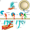
Model of how Munc18 prepares syntaxin for SNARE complex formation. A, schematic showing that Munc18 binds first to the closed conformation and N-peptide region of syntaxin. Munc18 then makes the bound syntaxin able to engage with its partner SNAREs, SNAP-25 and synaptobrevin. This step is triggered by additional factors, such as Munc13 (5), and possibly involves a conformational change of the helical hairpin region of Munc18. The crystal structure of Munc18 (cyan) bound to Syx1a (Habc domain (orange), SNARE domain (red)) is shown at the top (10). The region shown below as a close-up image (B and C) is indicated by a dashed square. Shown is a close-up of the different conformations of the helical hairpin region of Munc18a in complex with the closed conformation of Syx1a (“furled”; B) or bound to the N-peptide of Syx4 (“extended”; C) (49).

References

    1. Cai H., Reinisch K., and Ferro-Novick S. (2007) Coats, tethers, Rabs, and SNAREs work together to mediate the intracellular destination of a transport vesicle. Dev. Cell 12, 671–682 - PubMed
    1. Südhof T. C., and Rothman J. E. (2009) Membrane fusion: grappling with SNARE and SM proteins. Science 323, 474–477 - PMC - PubMed
    1. Jahn R., and Fasshauer D. (2012) Molecular machines governing exocytosis of synaptic vesicles. Nature 490, 201–207 - PMC - PubMed
    1. Hong W., and Lev S. (2014) Tethering the assembly of SNARE complexes. Trends Cell Biol. 24, 35–43 - PubMed
    1. Rizo J., and Xu J. (2015) The synaptic vesicle release machinery. Annu. Rev. Biophys. 44, 339–367 - PubMed

Publication types

MeSH terms

LinkOut - more resources