. 2018 Jan 1;119(1):124-133.
doi: 10.1152/jn.00958.2016. Epub 2017 Oct 18.

Path perturbation detection tasks reduce MSTd neuronal self-movement heading responses

Affiliations

Path perturbation detection tasks reduce MSTd neuronal self-movement heading responses

William K Page et al. J Neurophysiol. .

Abstract

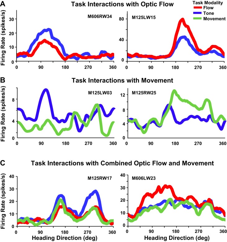
We presented optic flow and real movement heading stimuli while recording MSTd neuronal activity. Monkeys were alternately engaged in three tasks: visual detection of optic flow heading perturbations, vestibular detection of real movement heading perturbations, and auditory detection of brief tones. Push-button RTs were fastest for tones and slower for visual and vestibular heading perturbations, suggesting that the tone detection task was easier. Neuronal heading selectivity was strongest during the tone detection task, and weaker during the visual and vestibular heading perturbation detection tasks. Heading selectivity was weaker during visual and vestibular path perturbation detection, despite our presented heading cues only in the visual and vestibular modalities. We conclude that focusing on the self-movement transients of path perturbation distracted the monkeys from their heading and reduced neuronal responsiveness to heading direction. NEW & NOTEWORTHY Heading analysis is critical for steering and navigation. We recorded the activity of monkey cortical heading neurons during naturalistic self-movement. When the monkeys were required to respond to transient changes in their path, neuronal responses to heading direction were diminished. This suggests that the need to respond to momentary path perturbations reduces your ability to process your heading direction.

Keywords: attention; cortex; extrastriate; optic flow; visual motion.


Figures

Fig. 1.
Fig. 1.
Vestibular and visual self-movement stimuli were presented during single neuron recordings. A: monkeys underwent circular path translational self-movement (red circle) either viewing an optic flow simulation of circular path self-movement on a room-sized sled, or experiencing sled movement on a circular path, or congruently combined optic flow and sled movement. The monkey was engaged in three push-button task conditions: brief path perturbations (green wavelet) from the optic flow-simulated circular path, the sled movement circular path, or a brief audible tone presented along the circular path. In all three cue modalities, the push-button cue was presented at one of eight locations around the circle. B: a table representing the seven stimulus × cue conditions studied. Three heading stimulus modalities conditions were optic flow alone, sled movement alone, and combined stimuli (abscissa). Push-button task cue modalities combined path perturbation in the presented heading stimulus plus an auditory tone detection task.
Fig. 2.
Fig. 2.
Comparison of the response times for the various stimulus and task conditions in the two monkeys. The response times (ordinate) for the two monkeys (A: M606; B: M125; C: both) with the optic flow cue (red), translational movement cue (green), and tone cue (blue) stimuli are shown for each of three stimulus conditions (abscissa): optic flow alone (left), real movement alone (middle), and both optic flow and real movement (right). The reaction times were shortest for the tone-cued trials and longest for the sled movement-cued trials.
Fig. 3.
Fig. 3.
Activity of an MSTd neuron (firing rate, ordinate) recorded during circular path stimulus presentation (10.5 s, abscissa). The ±750-ms interval of cueing by path perturbations or audible tones was excluded from the records of each trial. A: activity evoked by optic flow circular path simulation is larger during the audible tone detection task (blue) than during the optic flow path perturbation detection task (red). B: activity evoked by translational movement circular path stimulation is similar during the audible tone detection task (blue) and the translational movement path perturbation detection task (green). C: activity evoked by congruently combined optic flow and translational movement circular path stimuli is largest during the audible tone detection task (blue), smaller during the optic flow path perturbation detection task (red), and greatly diminished during translational movement path perturbation detection task (green).
Fig. 4.
Fig. 4.
Task effects on heading-selective responses to circular path stimuli. A: neuronal activity (ordinate) throughout the optic flow circular path simulation (10.5 s, abscissa) during push-button cueing by optic flow path perturbation (red) or by an audible tone (blue). Left: a neuron that shows larger optic flow heading responses during the tone detection task. Right: a neuron that shows larger optic flow heading responses during the optic flow path perturbation detection task. B: task effects on heading-selective responses to sled movement circular path stimuli (format as in A). Left: a neuron that shows larger sled movement heading responses during the tone detection task. Right: a neuron that shows larger sled movement heading responses during the sled movement path perturbation detection task. C: task effects on heading-selective responses to combined optic flow and translational movement circular path stimuli (format as in A). Left: a neuron that shows a shift of heading selectivity with a larger response during the tone detection task. Right: a neuron that shows larger heading responses during the optic flow path perturbation detection task.
Fig. 5.
Fig. 5.
The bimodality of response profiles in the sample of neurons is illustrated for the three stimulus conditions: optic flow alone (A), sled movement alone (B), and combined stimuli (C). Left: neurons were split into two groups depending on whether the second peak was close (solid lines) or further away (dashed lines) from the preferred stimulus direction. The neurons with a distant second peak have a distinct second peak for all conditions. Right: scatterplots of the degree of unimodal selectivity (abscissa) vs. bimodal selectivity (ordinate) from Rayleigh z scores for the three stimulus conditions (A–C). There is a broad continuum of the relative strengths of unimodality and bimodality in all conditions. Illustrative examples are shown in the inset responses, and their position in the scatterplots is indicated by the nearer stars.
Fig. 6.
Fig. 6.
Effects of cue modality (line colors) on neuronal population-averaged responses (ordinates) across heading directions (abscissas). A: averaged population responses for heading directions simulated by optic flow do not show significant differences between the optic flow path perturbation detection task vs. the audible tone detection task. B: averaged population responses for heading directions in translational movement do not show significant differences between the translational movement path perturbation detection task vs. the audible tone detection task. C: averaged population responses for heading directions in combined optic flow and translational movement show significantly greater heading selectivity during the audible tone detection task than during the optic flow or translational movement path perturbation detection task. D: scatterplot of differences in averaged single neuron activity in the tone detection task minus the translational movement path perturbation detection task (ordinate), vs. in the tone detection task minus the optic flow path perturbation detection task (abscissa). The comparability of effects in the tone detection task, compared with the translational movement and optic flow path perturbation detection tasks, show similar degrees of task modulation in the visual and vestibular domains.

Similar articles

Cited by

References

    1. Angelaki DE, Gu Y, Deangelis GC. Visual and vestibular cue integration for heading perception in extrastriate visual cortex. J Physiol 589: 825–833, 2011. doi:10.1113/jphysiol.2010.194720. - DOI - PMC - PubMed
    1. Bremmer F, Kubischik M, Pekel M, Hoffmann KP, Lappe M. Visual selectivity for heading in monkey area MST. Exp Brain Res 200: 51–60, 2010. doi:10.1007/s00221-009-1990-3. - DOI - PubMed
    1. Chen A, DeAngelis GC, Angelaki DE. A comparison of vestibular spatiotemporal tuning in macaque parietoinsular vestibular cortex, ventral intraparietal area, and medial superior temporal area. J Neurosci 31: 3082–3094, 2011. doi:10.1523/JNEUROSCI.4476-10.2011. - DOI - PMC - PubMed
    1. Crist CF, Yamasaki DS, Komatsu H, Wurtz RH. A grid system and a microsyringe for single cell recording. J Neurosci Methods 26: 117–122, 1988. doi:10.1016/0165-0270(88)90160-4. - DOI - PubMed
    1. Crowell JA, Banks MS. Perceiving heading with different retinal regions and types of optic flow. Percept Psychophys 53: 325–337, 1993. doi:10.3758/BF03205187. - DOI - PubMed

Publication types

LinkOut - more resources