

On Myelinated Axon Plasticity and Neuronal Circuit Formation and Function
- PMID: 29046438
- PMCID: PMC6596541
- DOI: 10.1523/JNEUROSCI.3185-16.2017
On Myelinated Axon Plasticity and Neuronal Circuit Formation and Function
Abstract
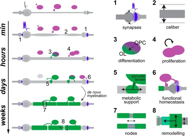
Studies of activity-driven nervous system plasticity have primarily focused on the gray matter. However, MRI-based imaging studies have shown that white matter, primarily composed of myelinated axons, can also be dynamically regulated by activity of the healthy brain. Myelination in the CNS is an ongoing process that starts around birth and continues throughout life. Myelin in the CNS is generated by oligodendrocytes and recent evidence has shown that many aspects of oligodendrocyte development and myelination can be modulated by extrinsic signals including neuronal activity. Because modulation of myelin can, in turn, affect several aspects of conduction, the concept has emerged that activity-regulated myelination represents an important form of nervous system plasticity. Here we review our increasing understanding of how neuronal activity regulates oligodendrocytes and myelinated axons in vivo, with a focus on the timing of relevant processes. We highlight the observations that neuronal activity can rapidly tune axonal diameter, promote re-entry of oligodendrocyte progenitor cells into the cell cycle, or drive their direct differentiation into oligodendrocytes. We suggest that activity-regulated myelin formation and remodeling that significantly change axonal conduction properties are most likely to occur over timescales of days to weeks. Finally, we propose that precise fine-tuning of conduction along already-myelinated axons may also be mediated by alterations to the axon itself. We conclude that future studies need to analyze activity-driven adaptations to both axons and their myelin sheaths to fully understand how myelinated axon plasticity contributes to neuronal circuit formation and function.
Keywords: axons; myelin; oligodendrocytes; plasticity.
Copyright © 2017 the authors 0270-6474/17/3710023-12$15.00/0.
Figures
References
Publication types
MeSH terms
Grants and funding
LinkOut - more resources
Full Text Sources
Other Literature Sources