. 2017 Dec 14;92(1):e01636-17.
doi: 10.1128/JVI.01636-17. Print 2018 Jan 1.

Distinct Roles of Cellular ESCRT-I and ESCRT-III Proteins in Efficient Entry and Egress of Budded Virions of Autographa californica Multiple Nucleopolyhedrovirus

Affiliations

Distinct Roles of Cellular ESCRT-I and ESCRT-III Proteins in Efficient Entry and Egress of Budded Virions of Autographa californica Multiple Nucleopolyhedrovirus

Qi Yue et al. J Virol. .

Abstract

The endosomal sorting complex required for transport (ESCRT) machinery is necessary for budding of many enveloped viruses. Recently, it was demonstrated that Vps4, the key regulator for recycling of the ESCRT-III complex, is required for efficient infection by the baculovirus Autographa californica multiple nucleopolyhedrovirus (AcMNPV). However, ESCRT assembly, regulation, and function are complex, and little is known regarding the details of participation of specific ESCRT complexes in AcMNPV infection. In this study, the core components of ESCRT-I (Tsg101 and Vps28) and ESCRT-III (Vps2B, Vps20, Vps24, Snf7, Vps46, and Vps60) were cloned from Spodoptera frugiperda Using a viral complementation system and RNA interference (RNAi) assays, we found that ESCRT-I and ESCRT-III complexes are required for efficient entry of AcMNPV into insect cells. In cells knocking down or overexpressing dominant negative (DN) forms of the components of ESCRT-I and ESCRT-III complexes, entering virions were partially trapped within the cytosol. To examine only egress, cells were transfected with the double-stranded RNA (dsRNA) targeting an individual ESCRT-I or ESCRT-III gene and viral bacmid DNA or viral bacmid DNA that expressed DN forms of ESCRT-I and ESCRT-III components. We found that ESCRT-III components (but not ESCRT-I components) are required for efficient nuclear egress of progeny nucleocapsids. In addition, we found that several baculovirus core or conserved proteins (Ac11, Ac76, Ac78, GP41, Ac93, Ac103, Ac142, and Ac146) interact with Vps4 and components of ESCRT-III. We propose that these viral proteins may form an "egress complex" that is involved in recruiting ESCRT-III components to a virus egress domain on the nuclear membrane.IMPORTANCE The ESCRT system is hijacked by many enveloped viruses to mediate budding and release. Recently, it was found that Vps4, the key regulator of the cellular ESCRT machinery, is necessary for efficient entry and egress of Autographa californica multiple nucleopolyhedrovirus (AcMNPV). However, little is known about the roles of specific ESCRT complexes in AcMNPV infection. In this study, we demonstrated that ESCRT-I and ESCRT-III complexes are required for efficient entry of AcMNPV into insect cells. The components of ESCRT-III (but not ESCRT-I) are also necessary for efficient nuclear egress of progeny nucleocapsids. Several baculovirus core or conserved proteins were found to interact with Vps4 and components of ESCRT-III, and these interactions may suggest the formation of an "egress complex" involved in the nuclear release or transport of viral nucleocapsids.

Keywords: AcMNPV; ESCRT-I; ESCRT-III; baculovirus; virus entry and egress.


Figures

FIG 1
FIG 1
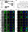
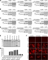
Construction and transient expression of GFP-tagged wild-type or truncated forms of ESCRT-I components Tsg101 and Vps28. (A) Schematic representation of the domain organization of WT Tsg101 and Vps28 and truncated forms of each. Numbers on the right denote the amino acid sequence length of each construct. Abbreviations: CC, coiled coil; CTD, C-terminal four-helix bundle domain; dUEV, deletion of UEV domain; PRD, proline-rich domain; SB, steadiness box; UEV, ubiquitin-enzyme variant domain. (B, C) Expression of GFP-tagged WT or truncated forms of Tsg101 and Vps28 in transfected Sf9 cells. (B) The expression of GFP-tagged Tsg101 and Vps28 constructs was analyzed by Western blotting using a GFP-specific polyclonal antibody; gels were spliced for labeling purposes. (C) The cellular distribution of GFP-tagged Tsg101 and Vps28 constructs was visualized by epifluorescence microscopy (Epi, left panels) and confocal microscopy (Confocal, right panels). Phase-contrast images on the left side illustrate the presence of vesicles induced by Vps28 construct Core, which lacks the CTD domain. (D) Colocalization of GFP-tagged Tsg101 and Vps28 constructs with mCherry-tagged Vps4 mutant E231Q in cotransfected Sf9 cells. Cell boundaries are traced with circular dashed lines. Bar, 10 μm.
FIG 2
FIG 2
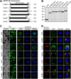
Transient expression of GFP-tagged ESCRT-III components in Sf9 cells. (A) Schematic representation of the ESCRT-III components cloned from Sf9 cells. The predicted Snf7 domain of each component is shown as a black box, and the start and end amino acids of Snf7 domains in individual components are indicated. The amino acid sequence length for each protein is indicated on the right. (B, C) Expression of GFP-tagged ESCRT-III proteins in transfected Sf9 cells. (B) The expression of GFP-tagged ESCRT-III proteins was analyzed by Western blotting using a GFP-specific polyclonal antibody; gels were spliced for labeling purposes. (C) The cellular distribution of GFP-tagged ESCRT-III proteins was visualized by epifluorescence microscopy (Epi, left panels) and confocal microscopy (Confocal, right panels). The presence of vesicles induced by DN ESCRT-III constructs can be observed in phase-contrast images on the left. (D) Colocalization of GFP-tagged ESCRT-III proteins with mCherry-tagged Vps4 mutant E231Q in cotransfected Sf9 cells. Cell boundaries are traced with circular dashed lines. Bar, 10 μm.
FIG 3
FIG 3
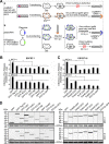
Transient expression of GFP-tagged ESCRT-I and ESCRT-III proteins significantly inhibits the production of infectious AcMNPV in a viral complementation assay. (A) Schematic representation of the viral complementation assay. (a). In cells transfected with a gp64 knockout AcMNPV bacmid, virus budding is defective. When the gp64 knockout bacmid DNA is transfected into Sf9Op1D cells that stably express OpMNPV GP64, virus budding and infectivity are complemented by OpMNPV GP64. (b) Sf9 cells are cotransfected with two plasmids separately expressing AcMNPV GP64 (pBieGP64) and GFP or a GFP-tagged ESCRT protein. At 16 h p.t., the cells are infected with a gp64 knockout AcMNPV virus that was produced in Sf9Op1D cells and containing the OpMNPV GP64 protein on its surface. Because all cells do not become transfected, the gp64 knockout AcMNPV can only bud and propagate in cells that are productively transfected, expressing both GFP or the GFP-tagged ESCRT protein and AcMNPV GP64, which complements the gp64 knockout. In nontransfected cells, the gp64 knockout AcMNPV virus can enter the cells, but budding of progeny virions is defective. (B, C) Sf9 cells were cotransfected with a plasmid expressing GP64 together with a plasmid encoding GFP-tagged ESCRT-I and ESCRT-III proteins, E231Q-GFP, or GFP. At 16 h p.t., the cells were infected with a gp64 knockout AcMNPV at an MOI of 1 or 5. At 24 h p.i., the titers of progeny viruses from cell culture medium were determined by TCID50 assay on a GP64-complementing cell line (Sf9OP1D). Error bars indicate the standard deviations from the means of results for triplicate samples. (D) The expressions of GP64 and GFP-tagged ESCRT-I and ESCRT-III proteins in cotransfected and infected cells were analyzed by Western blotting using anti-GP64 MAb (AcV5) and an anti-GFP polyclonal antibody; gels were spliced for labeling purposes. *, P < 0.005 (by unpaired t test).
FIG 4
FIG 4
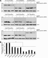
RNAi knockdown of ESCRT-I or ESCRT-III proteins inhibits production of infectious AcMNPV. (A) Sf9 cells were transfected with a plasmid expressing HA- or c-Myc-tagged ESCRT-I and ESCRT-III proteins or Vps4 or were cotransfected with a plasmid expressing individual HA- or c-Myc-tagged ESCRT protein and a dsRNA specific for an ESCRT gene or GFP. At 48 h p.t., the transfected cells were collected and expression of the HA- or c-Myc-tagged ESCRT protein was detected by Western blotting with an anti-HA monoclonal antibody or an anti-Myc polyclonal antibody. Actin was detected (using anti-β-actin) as a loading control. (B) Sf9 cells were mock transfected or transfected with the dsRNA specific for GFP or for an individual ESCRT gene. At 48 h p.t., the transfected cells were infected with control AcMNPV. At 24 h p.i., the cell culture supernatants were collected and virus titers were determined by TCID50. **, P < 0.0005 (by unpaired t test).
FIG 5
FIG 5
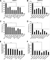
Effects of overexpression of GFP-tagged ESCRT-I and ESCRT-III proteins on early stages of AcMNPV replication. Sf9 cells were cotransfected with two plasmids separately expressing (i) GP64 and (ii) one of the GFP-tagged ESCRT-I or ESCRT-III proteins, E231Q-GFP, or GFP. (A, B) At 16 h p.t., the cells were infected with a gp64 knockout virus, LacZGUS-gp64ko (MOI = 5). At 6 h p.i., the infected cells were collected and the early reporter (beta-galactosidase) activity was measured using CPRG as the substrate. (C to F) At 24 h p.i., the parallel transfected and infected cells were lysed and the late reporter (GUS) activity was measured (C, D) and viral genomic DNA replication efficiency was evaluated by real-time PCR (E, F). Error bars indicate standard deviations of the means from three replicates. *, P < 0.005; **, P < 0.0005; ***, P < 0.00005 (by unpaired t test).
FIG 6
FIG 6
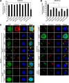
Analysis of the effects of overexpression of GFP-tagged ESCRT-I and ESCRT-III proteins on entry of AcMNPV. Sf9 cells were cotransfected with two plasmids separately expressing (i) GP64 and (ii) one of the GFP-tagged ESCRT-I or ESCRT-III proteins, E231Q-GFP, or GFP. At 16 h p.t., cells were infected with prechilled control AcMNPV or an mCherry-labeled AcMNPV virus (3mC) (MOI = 10 TCID50) at 4°C for 1 h, and then the cells were incubated at 27°C for 90 min. The control AcMNPV-infected cells were lysed, and the internalized viral genomic DNAs were determined by real-time PCR (A, B). The 3mC virus-infected cells were fixed and analyzed by confocal microscopy (C). Cell boundaries are traced with circular dashed lines. Bar, 10 μm. *, P < 0.005; **, P < 0.0005 (by unpaired t test).
FIG 7
FIG 7
Analysis of the effects of RNAi knockdowns targeting specific ESCRT-I and ESCRT-III genes on entry of AcMNPV. Sf9 cells were mock transfected or transfected with the dsRNA targeting an individual ESCRT-I or ESCRT-III gene, Vps4, or GFP. At 48 h p.t., cells were infected with prechilled control AcMNPV or an mCherry-labeled AcMNPV virus (3mC) (MOI = 10) at 4°C for 1 h, and then the cells were incubated at 27°C for 90 min. The control AcMNPV-infected cells were lysed, and the internalized viral genomic DNAs were determined by quantitative real-time PCR (A). The 3mC virus infected cells were fixed and analyzed by confocal microscopy (B). Cell boundaries are traced with dashed lines. Bar, 10 μm. Error bars represent standard deviations from the means of results from three replicates. **, P < 0.0005 (by unpaired t test).
FIG 8
FIG 8
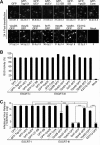
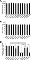
Infectious BV production in the presence of GFP-tagged ESCRT-I and ESCRT-III proteins expressed from AcMNPV bacmids. Sf9 cells were transfected with AcMNPV bacmids expressing either (i) one of the GFP-tagged ESCRT-I or ESCRT-III proteins, (ii) E231Q-GFP, or (iii) GFP. At 24 h p.t., the percentage of GFP-expressing cells was determined for each treatment, and percentages are shown below each panel as an estimate of transfection efficiency (A). A parallel group of transfected cells were also lysed at 24 h p.t., and GUS activity (expressed from late GUS reporter gene, driven by a p6.9 late promoter in each bacmid) was determined (B). The production of infectious BV from each treatment was determined by TCID50 assay of the cell supernatant (C). Error bars represent standard deviations from the means for three replicates. *, P < 0.005; ***, P < 0.00005 (by unpaired t test).
FIG 9
FIG 9
Analysis of the effects of RNAi knockdowns targeting specific ESCRT-I and ESCRT-III genes on infectious AcMNPV BV release. Sf9 cells were mock transfected or transfected with the dsRNA targeting an individual ESCRT-I or ESCRT-III gene or the control GFP gene. At 48 h p.t., the cells were transfected again with control AcMNPV bacmid DNA (AcMNPV-LacZGUS). After transfection with the viral bacmid DNA for 24 h, the transfected cells were lysed, and beta-Gal and GUS activities (separately expressed from early LacZ and late GUS reporter genes, driven by an ie2 early promoter and a p6.9 late promoter, respectively, in each bacmid) were determined (A, B). The production of infectious BV from each treatment was determined by TCID50 assay of the cell supernatant (C). Error bars represent standard deviations from the means for three replicates. *, P < 0.005; ***, P < 0.00005 (by unpaired t test).
FIG 10
FIG 10
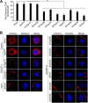
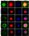
DN ESCRT-III and Vps4 proteins appear to inhibit the nuclear release of nucleocapsids. Sf9 cells were transfected with AcMNPV bacmids expressing VP39-mCherry and either Vps24-GFP, Snf7-GFP, Vps60-GFP, E231Q-GFP, or the control GFP. At 24 h p.t., the transfected cells were fixed and analyzed by confocal microscopy. Bar, 10 μm.
FIG 11
FIG 11
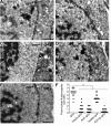
TEM analysis of Sf9 cells transfected with AcMNPV bacmid DNAs expressing DN ESCRT-III and Vps4 proteins. (A to E) Sf9 cells were transfected with AcMNPV bacmids expressing VP39-mCherry and either GFP (A), Vps24-GFP (B), Snf7-GFP (C), Vps60-GFP (D), or E231Q-GFP (E). At 72 h p.t., the transfected cells were fixed and analyzed by transmission electron microscopy. The nuclear membrane (nm), cytoplasmic membrane (cm), and nucleocapsids (white arrows) are indicated. Multiple aggregated nucleocapsids localized in the space between the inner and outer nuclear membrane are indicated by closed triangles. (F) The numbers of postnuclear nucleocapsids were determined, and these include those residing in the cytoplasm and budding through the cytoplasmic membrane. Numbers were calculated from 13 cells for each construct. Bar, 1 μm. **, P < 0.0005 (by unpaired t test).
FIG 12
FIG 12
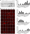
BiFC analysis of the interaction of ESCRT-III and AcMNPV proteins. (A) Sf9 cells were transfected with plasmids expressing each construct consisting of the N- or C-terminal domain of mCherry (Nm and Cm) fused with each ESCRT-III or viral protein. At 36 h p.t., the expression of each fusion protein construct was analyzed by Western blotting with an anti-HA MAb (ESCRT-III proteins) or an anti-Myc polyclonal antibody (viral proteins); gels were spliced for labeling purposes. (B) Fluorescence complementation in cells expressing Nm and Cm fused with ESCRT-III and viral proteins. Sf9 cells were cotransfected with two plasmids separately expressing Nm or Cm fused with ESCRT-III or viral proteins. At 36 h p.t., the cells were photographed using epifluorescence microscopy. Labels on the left and top identify the cotransfected construct pairs in each panel. Cell boundaries are traced with circular dashed lines. (C) The bar graphs show the percentages of mCherry-positive cells in cotransfected Sf9 cells expressing Nm- and Cm-fused ESCRT-III and viral proteins. The pairs of cotransfected constructs are indicated below the x axis of each graph. Error bars represent standard deviations from the means for three replicates.
FIG 13
FIG 13
Coimmunoprecipitation and BiFC analysis of interactions of Vps4 and AcMNPV proteins. (A to D) Sf9 cells were transfected with the indicated (+ or −) plasmids or combinations of plasmids expressing either HA-tagged viral proteins or Myc-tagged Vps4 or modified Vps4 constructs (E231Q and K176Q). At 36 h p.t., the transfected and cotransfected cells were separately lysed and subjected to immunoprecipitation with anti-HA monoclonal antibodies and protein G agarose. The precipitates (Co-IP) were detected on Western blots with an anti-Myc polyclonal antibody (right panel in each group). The cell lysates (Lysate) were also examined on Western blots with an anti-HA monoclonal antibody (top panels) or an anti-Myc polyclonal antibody (bottom panels). Abs, antibodies. (E) Sf9 cells were transfected with a plasmid expressing the N- or C-terminal domain of mCherry (Nm and Cm) fused with Vps4, Vps4 with DN mutations (E231Q and K176Q), or viral proteins (Ac11, Ac93, Ac103, or GP41). At 36 h p.t., expression of the fusion proteins was analyzed by Western blotting using anti-HA MAb (Vps4 and its DN mutations K176Q and E231Q) or an anti-Myc polyclonal antibody (viral proteins); gels were spliced for labeling purposes. (F) BiFC analysis of cells coexpressing Vps4 and viral protein pairs. Sf9 cells were cotransfected with two plasmids: one that expressed Nm-fused Vps4, E231Q, or K176Q and a second plasmid that expressed Cm-fused viral proteins Ac11, Ac93, Ac103, or GP41. At 36 h p.t., the cells were photographed using epifluorescence microscopy. Labels on the left and top identify the cotransfected construct pairs in each panel. Cell boundaries are traced with circular dashed lines. (G) The bar graphs show the percentages of mCherry-positive cells in cotransfected Sf9 cells expressing Nm- and Cm-fused Vps4 and viral proteins. The pairs of cotransfected constructs are indicated below the x axis of each graph. Error bars represent standard deviations from the means for three replicates.
FIG 14
FIG 14
BiFC analysis of interactions of AcMNPV proteins. (A) Sf9 cells were transfected with plasmids expressing the N- or C-terminal domain of mCherry (Nm or Cm) fused to viral proteins (Ac11, Ac76, Ac78, Ac93, Ac103, Ac146, and GP41). At 36 h p.t., the expression of each fusion protein was analyzed by Western blotting using an anti-HA MAb (Nm-fused viral proteins) or an anti-Myc polyclonal antibody (Cm-fused viral proteins) for detection; gels were spliced for labeling purposes. (B) BiFC analysis of cells coexpressing Nm- and Cm-fused viral proteins. Sf9 cells were cotransfected with two plasmids, separately expressing Nm- or Cm-fused viral proteins. The pairs of cotransfected constructs are indicated at the top and left of each panel. At 36 h p.t., cells were photographed using epifluorescence microscopy and analyzed. Cell boundaries are traced with circular dashed lines. (C) Percentages of mCherry-positive cells in transfected Sf9 cells expressing Nm- and Cm-fused viral proteins. The pairs of cotransfected constructs are indicated below the x axis of each graph. Error bars represent standard deviations from the means for three replicates.
FIG 15
FIG 15
Schematic representation of the protein-protein interaction network of ESCRT-III proteins and Vps4 and viral proteins and ESCRT-III/Vps4. ESCRT-III components and viral proteins that interact with themselves are shown as shaded circles. The top left panel shows interactions among ESCRT-III proteins (Vps2B, Vps20, Vps24, Vps46, Vps60, and Snf7) and Vps4. The panel on the right shows interactions among the viral proteins (inner circle, Ac11, Ac76, Ac78, Ac93, Ac103, Ac142, Ac146, and GP41) and interactions between each viral protein (inner group) and ESCRT-III proteins (outer group, Vps2B, Vps20, Vps24, Vps46, Vps60, and Snf7). The lower left panel shows interactions between cellular Vps4 and viral proteins (Ac93, Ac103, and GP41).
FIG 16
FIG 16
A hypothetical model of the interaction of the viral proteins and ESCRT-III/Vps4 in nuclear egress of progeny nucleocapsids. (A) In AcMNPV-infected cells, the nuclear membrane-associated Ac76 may initiate the nuclear membrane protrusion. Ac76 interacts with Ac93 and Ac78, which may form a complex that interacts with Ac103, which in turn interacts with nucleocapsid-associated protein Ac146, to target the progeny nucleocapsids to the budding region on the nuclear membrane. (B) A viral protein complex (Ac76, Ac93, Ac78, and possibly Ac142) may recruit the core components of ESCRT-III to the budding region and result in the formation of the Snf7 filament that constricts the nuclear membrane, releasing a double-membraned vesicle containing nucleocapsids. (C) After pinching off the double-membraned vesicle, the viral protein complex within the nucleus recruits Vps4 and its regulatory ESCRT-III proteins (Vps46 and Vps60) to form the activated Vps4 complex, which disassembles and recycles the ESCRT-III complex. BV, budded virions; CCV, clathrin-coated vesicle; DN, dominant negative; EE, early endosome; ER, endoplasmic reticulum; INM, inner nuclear membrane; LE/MVBs, late endosome/multivesicular bodies; NPC, nuclear pore complex; ONM, outer nuclear membrane; VS, virogenic stroma.

Similar articles

Cited by

References

    1. Henne WM, Buchkovich NJ, Emr SD. 2011. The ESCRT pathway. Dev Cell 21:77–91. doi:10.1016/j.devcel.2011.05.015. - DOI - PubMed
    1. Hurley JH. 2015. ESCRTs are everywhere. EMBO J 34:2398–2407. doi:10.15252/embj.201592484. - DOI - PMC - PubMed
    1. Guizetti J, Gerlich DW. 2012. ESCRT-III polymers in membrane neck constriction. Trends Cell Biol 22:133–140. doi:10.1016/j.tcb.2011.11.007. - DOI - PubMed
    1. Henne WM, Stenmark H, Emr SD. 2013. Molecular mechanisms of the membrane sculpting ESCRT pathway. Cold Spring Harb Perspect Biol 5(9):a016766. doi:10.1101/cshperspect.a016766. - DOI - PMC - PubMed
    1. Webster BM, Colombi P, Jager J, Lusk CP. 2014. Surveillance of nuclear pore complex assembly by ESCRT-III/Vps4. Cell 159:388–401. doi:10.1016/j.cell.2014.09.012. - DOI - PMC - PubMed

MeSH terms

Substances