. 2017 Oct 19;2(20):e94375.
doi: 10.1172/jci.insight.94375.

Human alternative Klotho mRNA is a nonsense-mediated mRNA decay target inefficiently spliced in renal disease

Affiliations

Human alternative Klotho mRNA is a nonsense-mediated mRNA decay target inefficiently spliced in renal disease

Rik Mencke et al. JCI Insight. .

Abstract

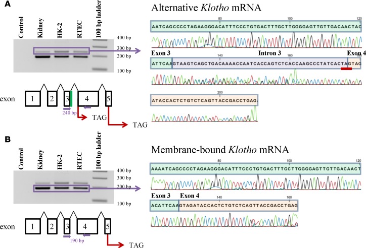
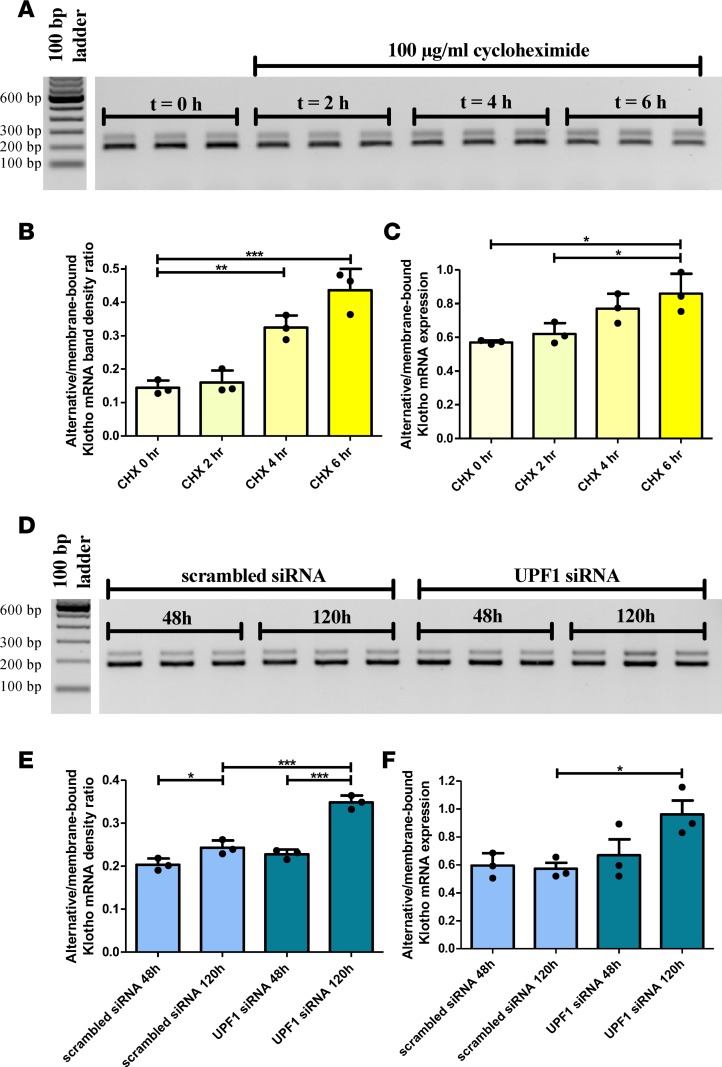
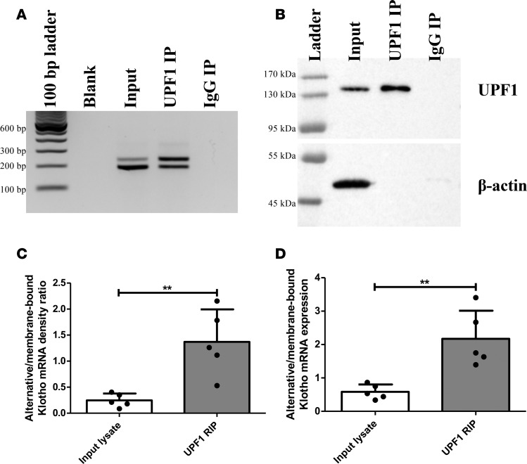
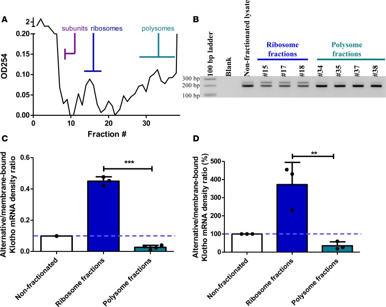
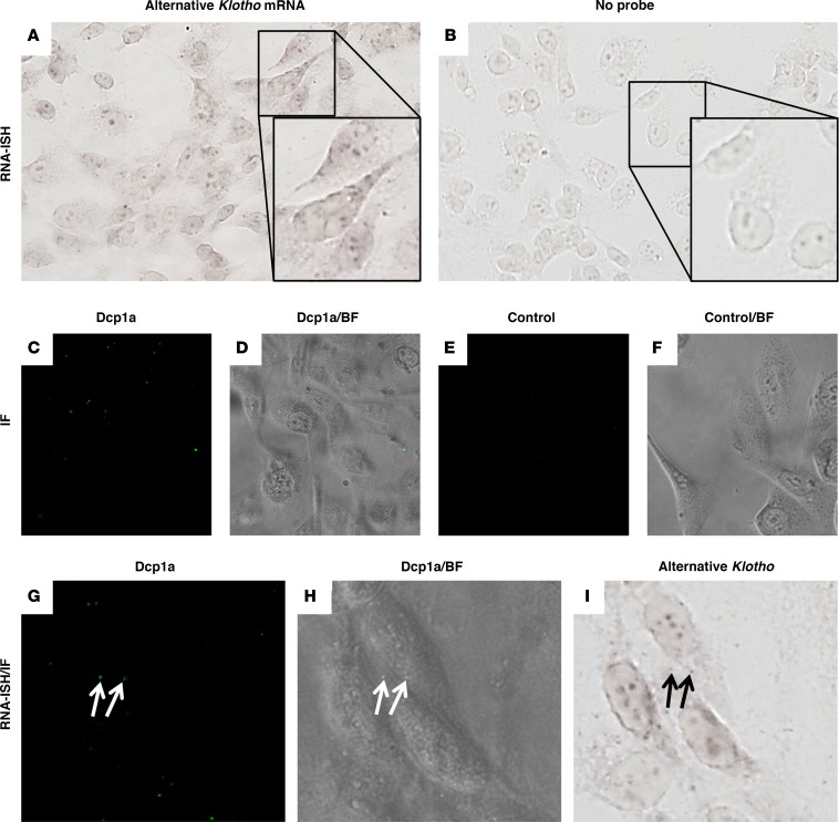
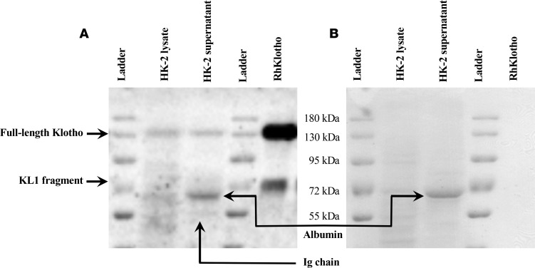
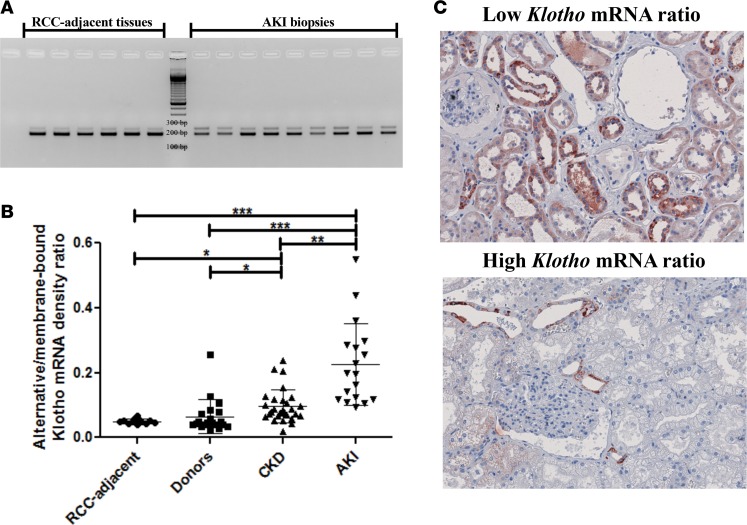
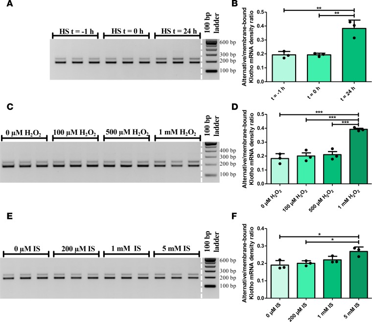
Klotho is a renal protein involved in phosphate homeostasis, which is downregulated in renal disease. It has long been considered an antiaging factor. Two Klotho gene transcripts are thought to encode membrane-bound and secreted Klotho. Indeed, soluble Klotho is detectable in bodily fluids, but the relative contributions of Klotho secretion and of membrane-bound Klotho shedding are unknown. Recent advances in RNA surveillance reveal that premature termination codons, as present in alternative Klotho mRNA (for secreted Klotho), prime mRNAs for degradation by nonsense-mediated mRNA decay (NMD). Disruption of NMD led to accumulation of alternative Klotho mRNA, indicative of normally continuous degradation. RNA IP for NMD core factor UPF1 resulted in enrichment for alternative Klotho mRNA, which was also not associated with polysomes, indicating no active protein translation. Alternative Klotho mRNA transcripts colocalized with some P bodies, where NMD transcripts are degraded. Moreover, we could not detect secreted Klotho in vitro. These results suggest that soluble Klotho is likely cleaved membrane-bound Klotho only. Furthermore, we found that, especially in acute kidney injury, splicing of the 2 mRNA transcripts is dysregulated, which was recapitulated by various noxious stimuli in vitro. This likely constitutes a novel mechanism resulting in the downregulation of membrane-bound Klotho.


Conflict of interest statement

Conflict of interest: The authors have declared that no conflict of interest exists.

Figures

Figure 1
Figure 1. Identification of human Klotho mRNA transcripts by RT-PCR and DNA sequencing.
(A) The alternative Klotho mRNA transcript is detected in human kidney, primary renal tubular epithelial cells, and in the human HK-2 renal cell line. PCR using primers spanning exons 3 and 4 (indicated by closed purple arrows) yields 2 PCR products. DNA sequencing confirms that the 240 bp PCR product corresponds to the alternative Klotho mRNA, which contains a 50 bp insertion (purple) between exon 3 (green) and 4 (orange), yielding a premature TAG stop codon (underlined in red). The alternative Klotho exons are depicted schematically, including the intronic sequence between exons 3 and 4 (green). (Note: nucleotide 145 in this sequence has similarly low signals for G and C, but is a G according to published sequences.) (B) The membrane-bound Klotho mRNA transcript is detected in human kidney, in primary renal tubular epithelial cells, and in the human HK-2 renal cell line. PCR using primers spanning exons 3 and 4 (indicated by closed purple arrows) yields 2 PCR products. DNA sequencing confirms that the 190 bp PCR product corresponds to the membrane-bound Klotho mRNA, which does not contain the 50 bp insertion between exon 3 and 4. The membrane-bound Klotho exons are depicted schematically.
Figure 2
Figure 2. Blocking nonsense-mediated mRNA decay, using cycloheximide or by silencing UPF1, results in the accumulation of the alternative Klotho mRNA transcript in HK-2 cells.
(A) RT-PCR for the 2 Klotho transcripts in HK-2 cells incubated with 100 μg/ml cycloheximide for 0, 2, 4, or 6 hours, showing accumulation of the alternative Klotho transcript. (B) Densitometric quantification of A, expressed as the ratio of the alternative and membrane-bound Klotho mRNAs (values are provided in Supplemental Table 1). (C) qPCR analysis for both Klotho transcripts, using ΔCt = Ct(alternative Klotho transcript) – Ct(membrane-bound Klotho transcript), which confirms the RT-PCR results. (D) RT-PCR for the 2 Klotho transcripts in HK-2 cells after Upf1 or scrambled siRNA transfection for 48 or 120 hours, showing accumulation of the alternative Klotho transcript after 120 hours. (E) Densitometric quantification of D, expressed as the ratio of the alternative and membrane-bound Klotho mRNAs (values are provided in Supplemental Table 2). (F) qPCR analysis for both Klotho transcripts, using ΔCt = Ct(alternative Klotho transcript) – Ct(membrane-bound Klotho transcript), confirms the RT-PCR results. *P < 0.05, **P < 0.01, ***P < 0.001, as tested by one-way ANOVA with post-hoc Bonferroni correction. All individual data points represent means of 3 independent experiments, performed in triplicate (plotted with mean ± SD).
Figure 3
Figure 3. RNA IP of HK-2 cell lysate reveals enrichment of the alternative Klotho mRNA associated with UPF1.
(A) RT-PCR analysis for both Klotho transcripts in HK-2 cell lysate (input), after IP for UPF1, and after IP with rabbit IgG, showing enrichment for the alternative Klotho mRNA in UPF1-associated RNA. (B) Western blot analysis for UPF1 and β-actin in HK-2 cell lysate and after IP with rabbit anti-UPF1 or rabbit IgG, showing enrichment for UPF1 and depletion of other proteins, as indicated by absence of β-actin in IP fractions. (C) Densitometric quantification of A, expressed as the ratio of the alternative and membrane-bound Klotho mRNAs. (D) qPCR analysis for both Klotho transcripts, using ΔCt = Ct(alternative Klotho transcript) – Ct(membrane-bound Klotho transcript), confirms the RT-PCR results. **P < 0.01, as tested by Student’s t test. All individual data points represent values from independent experiments (with mean ± SD).
Figure 4
Figure 4. Polysome fractionation of HK-2 cell lysate on sucrose gradients reveals enrichment of the alternative Klotho mRNA associated with single ribosomes and depletion in polysome-associated fractions.
(A) Representative absorbance spectrum of sucrose gradient fractions at 254 nm allows for identification of fractions enriched for free material, single ribosome subunits, single ribosomes, and polysomes. (B) RT-PCR analysis for both Klotho transcripts in nonfractionated HK-2 cell lysate, in single ribosome–associated fractions, and in polysome-associated fractions, showing enrichment for the alternative Klotho mRNA on single ribosomes and depletion in polysomes. (C) Densitometric quantification of the experiment in B, expressed as the ratio of the alternative and membrane-bound Klotho mRNAs. Individual data points refer to individual fractions (with mean ± SD). (D) Overall densitometric quantification of 3 replicate experiments, expressed as the ratio of the alternative and membrane-bound Klotho mRNAs, standardized to the nonfractionated lysate. **P < 0.01, ***P < 0.001, as tested by Student’s t test. Individual data points represent means of independent experiments with averages of 2–4 fractions in the same range (with means ± SD).
Figure 5
Figure 5. RNA in situ hybdridization for the alternative Klotho mRNA transcript in HK-2 cells reveals colocalization with some P bodies.
(A) RNA in situ hybridization for the alternative Klotho mRNA transcript indicates expression in HK-2 cells. (B) Control cells hybridized without probe. Insets magnify individual cells. (C) Immunofluorescence for Dcp1a identifies P bodies in HK-2 cells, (D) merged with bright-field image. (E) Immunofluorescence without primary antibody, (F) merged with bright-field image. (G) Immunofluorescence for Dcp1a in HK-2 cells, (H) merged with bright-field image. (I) RNA ISH reveals that some P bodies in the HK-2 cells in G and H colocalize with the signal for the alternative Klotho mRNA transcript (arrows). Images depicted in panels A, B, and I were digitally contrast-enhanced to 150% in Microsoft Powerpoint. Original magnifications are 400×; insets are 2-fold the magnification of A and B.
Figure 6
Figure 6. A putative secreted Klotho protein is not detected in HK-2 cell supernatant.
(A) Western blot for Klotho on HK-2 cell lysate, 48 hour–conditioned supernatant, and recombinant human (Rh) Klotho. Note the 130 kDa bands corresponding to full-length Klotho in HK-2 cells and in supernatant, similar to 130 kDa positive control recombinant Klotho. While a 70 kDa KL1 band was detected in the positive control, no such band was detected in HK-2 cell supernatant. Note that the relative densities of the recombinant protein and KL1 fragment are unrelated to the expected secondary cleavage of 130 kDa soluble Klotho generated by HK-2 cells. Smaller bands were determined to correspond to albumin and a likely immunoglobulin chain, as detailed in Supplemental Figure 3. (B) Ponceau S protein staining showing a prominent band at 65 kDa in HK-2 supernatant, corresponding to albumin, which is likely residual BSA from FCS. This band is further characterized in Supplemental Figure 3.
Figure 7
Figure 7. The relative abundance of the Klotho gene transcripts is dysregulated in vivo in renal disease.
(A) RT-PCR analysis of healthy renal cortices and acute kidney injury samples for both Klotho transcripts. (B) Densitometric quantification of renal cell carcinoma–adjacent (RCC-adjacent) healthy renal cortices (n = 12), donor kidney biopsies (n = 20), biopsies from kidneys afflicted with chronic kidney disease (CKD) (n = 28), and biopsies from kidneys suffering from acute kidney injury (AKI) (n = 18). (C) IHC for Klotho on kidneys with high alternative/membrane-bound Klotho mRNA ratios (n = 3) and kidneys with low alternative/membrane-bound Klotho mRNA ratios (n = 3) reveals lower Klotho protein expression in the kidneys with a relatively higher alternative Klotho mRNA abundance. Original magnifications: 200×. *P < 0.05, **P < 0.01, ***P < 0.001, as tested by Kruskal-Wallis test with Dunn’s post-hoc correction. Individual data points are plotted with mean ± SD.
Figure 8
Figure 8. The relative abundance of the Klotho gene transcripts is dysregulated in vitro after different stimuli.
(A) RT-PCR analysis for both Klotho transcripts after application of a heat shock as a physical stressor (45 min at 43°C) to HK-2 cells, that were lysed before the heat shock (t = –1 hour), after the heat shock (t = 0 hour), or 24 hours later. (B) Densitometric quantification of A, showing an increase in the alternative/membrane-bound Klotho mRNA ratio after 24 hours. (C) RT-PCR analysis for both Klotho transcripts after stimulation with different concentrations of H2O2 for 24 hours, to induce oxidative stress in HK-2 cells. (D) Densitometric quantification of C, showing an increase in the alternative/membrane-bound Klotho mRNA ratio after stimulation. (E) RT-PCR analysis for both Klotho transcripts after stimulation of HK-2 cells with different concentrations of indoxyl sulfate for 48 hours, as a model for uremia. (F) Densitometric quantification of E, showing a dose-dependent increase in the alternative/membrane-bound Klotho mRNA ratio after stimulation. Depicted are individual data points representing mean of 3 independent experiments, performed in triplicate (plotted with mean ± SD). *P < 0.05, **P < 0.01, ***P < 0.001, as tested by one-way ANOVA with Bonferroni’s post-hoc test.

Similar articles

Cited by

References

    1. Kuro-o M, et al. Mutation of the mouse klotho gene leads to a syndrome resembling ageing. Nature. 1997;390(6655):45–51. doi: 10.1038/36285. - DOI - PubMed
    1. Kurosu H, et al. Suppression of aging in mice by the hormone Klotho. Science. 2005;309(5742):1829–1833. doi: 10.1126/science.1112766. - DOI - PMC - PubMed
    1. Zhou L, Li Y, Zhou D, Tan RJ, Liu Y. Loss of Klotho contributes to kidney injury by derepression of Wnt/β-catenin signaling. J Am Soc Nephrol. 2013;24(5):771–785. doi: 10.1681/ASN.2012080865. - DOI - PMC - PubMed
    1. Hu MC, et al. Klotho and phosphate are modulators of pathologic uremic cardiac remodeling. J Am Soc Nephrol. 2015;26(6):1290–1302. doi: 10.1681/ASN.2014050465. - DOI - PMC - PubMed
    1. Dubal DB, et al. Life extension factor klotho prevents mortality and enhances cognition in hAPP transgenic mice. J Neurosci. 2015;35(6):2358–2371. doi: 10.1523/JNEUROSCI.5791-12.2015. - DOI - PMC - PubMed

Publication types