. 2017 Oct 19;2(20):e87322.
doi: 10.1172/jci.insight.87322.

Tracking mesenchymal stem cell contributions to regeneration in an immunocompetent cartilage regeneration model

Affiliations

Tracking mesenchymal stem cell contributions to regeneration in an immunocompetent cartilage regeneration model

Daniela Zwolanek et al. JCI Insight. .

Abstract

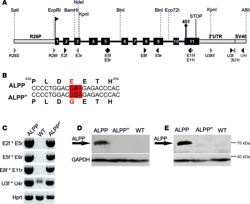
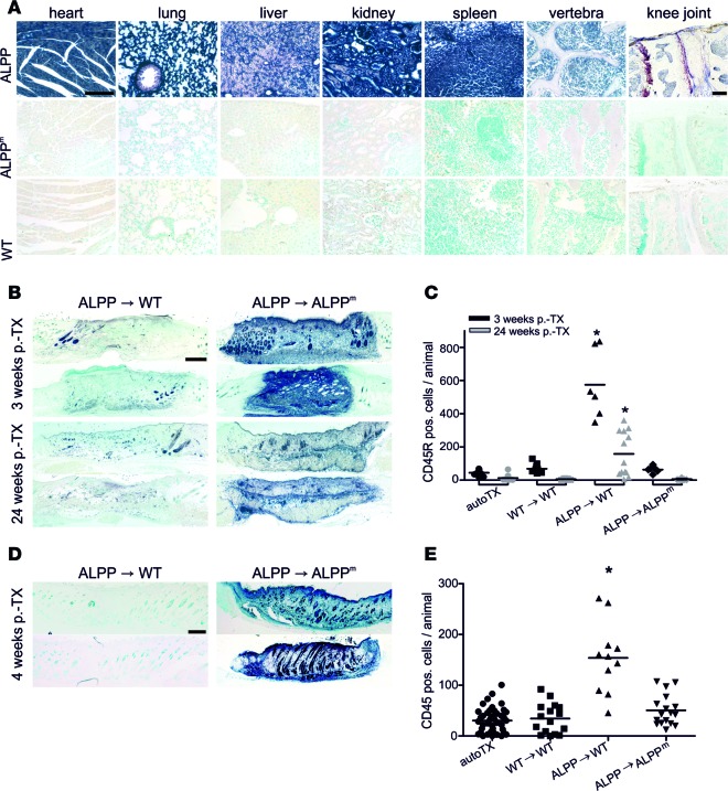
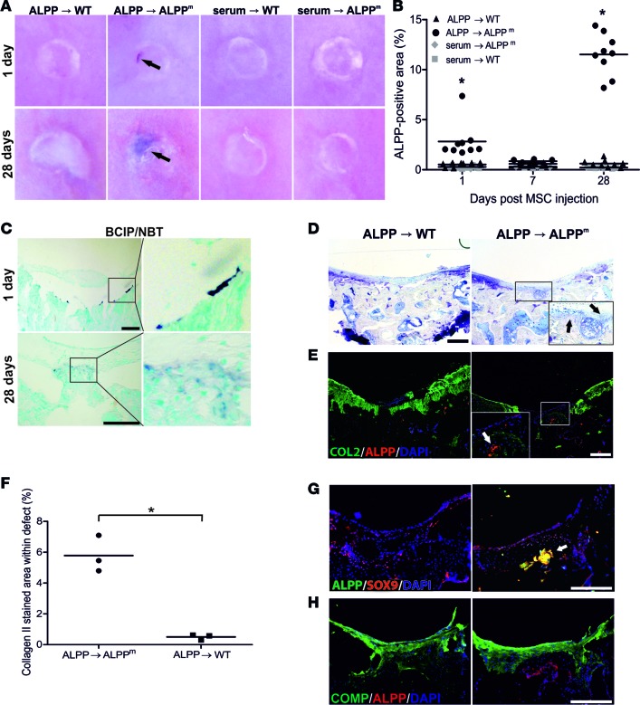
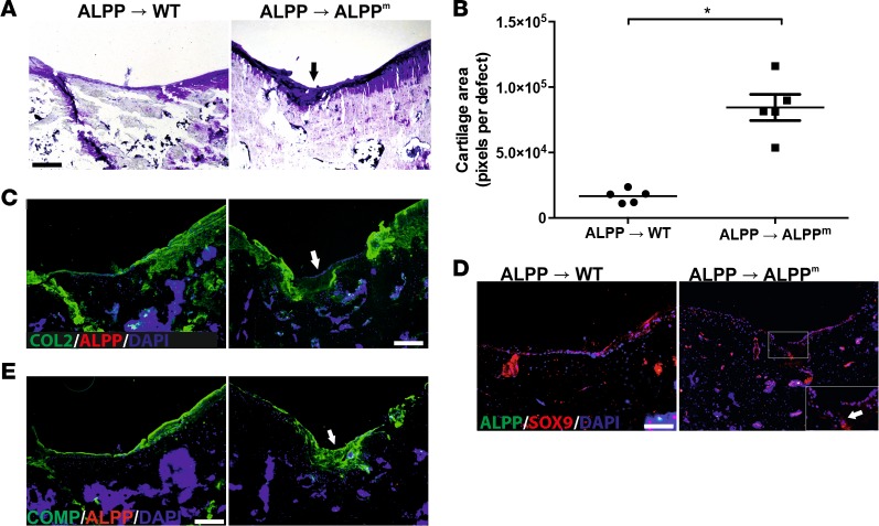
It is currently controversially discussed whether mesenchymal stem cells (MSC) facilitate cartilage regeneration in vivo by a progenitor- or a nonprogenitor-mediated mechanism. Here, we describe a potentially novel unbiased in vivo cell tracking system based on transgenic donor and corresponding immunocompetent marker-tolerant recipient mouse and rat lines in inbred genetic backgrounds. Tolerance of recipients was achieved by transgenic expression of an immunologically neutral but physicochemically distinguishable variant of the marker human placental alkaline phosphatase (ALPP). In this dual transgenic system, donor lines ubiquitously express WT, heat-resistant ALPP protein, whereas recipient lines express a heat-labile ALPP mutant (ALPPE451G) resulting from a single amino acid substitution. Tolerance of recipient lines to ALPP-expressing cells and tissues was verified by skin transplantation. Using this model, we show that intraarticularly injected MSC contribute to regeneration of articular cartilage in full-thickness cartilage defects mainly via a nonprogenitor-mediated mechanism.


Conflict of interest statement

Conflict of interest: The authors have declared that no conflict of interest exists.

Figures

Figure 1
Figure 1. Structure of human placental alkaline phosphatase (ALPP) transgenic constructs and transgene expression in transgenic mouse and rat lines.
(A) Structure of transgenic constructs for pronuclear injection, including relevant endonuclease restriction sites and primer positions. A point mutation in codon 451 of the ALPP gene introduced by site-directed mutagenesis results in an E451G substitution, giving rise to the heat-labile ALPPE451G (ALPPm) derivative. (B) Variation in amino acid sequence of heat-stable WT ALPP and heat-labile ALPPm. (C) Transcription of full-length WT and mutated ALPP was verified by RT-PCR on lung cDNA with amplicons spanning the whole transcript, exemplarily shown for the mouse model. n ≥ 7 per genotype. (D and E) Protein expression was examined by immunoblotting of mouse lung (D, n = 4 per genotype) and rat kidney tissue (E, n = 4 per genotype), showing high ALPP expression in ALPP-transgenic donor lines but no ALPP/ALPPm protein expression in ALPPm-transgenic and WT mice. GAPDH was used as loading control.
Figure 2
Figure 2. Histochemical ALPP staining and long-term tolerance of Tg(ALPPm) recipients to skin grafts from Tg(ALPP) donors.
(A) ALPP histochemistry on paraffin or plastic (bone and knee joint) sections from various organs of Tg(ALPP), Tg(ALPPm), and WT mice after a 35-minute heat inactivation at 72°C. Tg(ALPP) donors show strong staining in all tissues, whereas no enzyme activity could be detected in WT and Tg(ALPPm) mice. Scale bar: 50 μm. n = 10 per group. (B) Histochemistry of skin grafts from Tg(ALPP) at 3 and 24 weeks after transplantation. Strong ALPP staining was present in Tg(ALPP) grafts transplanted into Tg(ALPPm) recipients. Grafts from Tg(ALPP) mice were rejected by WT recipients, shown by the pronounced decline in ALPP staining within the grafts over time. Scale bar: 500 μm. n = 10 per group. (C) Leukocyte infiltration of skin grafts from Tg(ALPP) donor mice was quantified by CD45R immunostaining at 3 and 24 weeks after surgery and expressed as number of positive cells per tissue section. n ≥ 5 per group. (D) Histochemistry of skin grafts from Tg(ALPP) rats at 4 weeks after transplantation. Strong ALPP staining was present in Tg(ALPP) grafts transplanted into Tg(ALPPm) recipients. Grafts from Tg(ALPP) rats were rejected by WT recipients, as shown by the almost absent ALPP staining within the grafts. Scale bar: 500 μm. n = 10 per group. (E) Leukocyte infiltration of skin grafts from Tg(ALPP) donor rats was quantified by CD45 immunostaining at 4 weeks after transplantation and expressed as number of positive cells per tissue section. n ≥ 10 per group. *P < 0.05 by one-way ANOVA followed by Student-Newman-Keuls multiple comparison test. ALPP, human placental alkaline phosphatase; ALPPm, ALPPE451G mutant; ALPP ALPPm, transplantation from Tg(ALPP) donor into Tg(ALPPm) recipient; TX, transplantation.
Figure 3
Figure 3. Intraarticularly injected MSC contribute to cartilage regeneration in a rat model of full-thickness cartilage defects.
(A) Histochemical ALPP staining of the distal femoral joint surface with a full-thickness cartilage defect in the patellar groove 1 and 28 days after intraarticular injection of serum or 1 × 10⁷ MSC from Tg(ALPP) donor rats. Arrows show ALPP-positive staining in the defects. ALPP staining was absent in WT or Tg(ALPPm) recipients injected with MSC from Tg(ALPP) donors or serum, respectively. Scale bar: 250 μm. (B) Quantification of the ALPP-positive area within the full-thickness defects. Dots represent ALPP-positive area per animal. n ≥ 5 per group. *P < 0.05, ALPP → ALPPm recipients vs. all other groups by Kruskal-Wallis test followed by Mann-Whitney U test. (C) Histochemical ALPP staining of cryosections from femoral full-thickness cartilage defects 1 and 28 days after injection of 1 × 10⁷ MSC from Tg(ALPP) donor rats into the knee of Tg(ALPPm) recipients. Insets show ALPP-positive cells at the bottom of the defects. Scale bar: 50 μm. n = 3 animals per group. (D) Toluidine blue staining of cryosections from full-thickness cartilage defects, 28 days after injection of 1 × 10⁷ MSC from Tg(ALPP) donor rats into WT or Tg(ALPPm) recipients. Black arrows indicate induction of neocartilage formation within the defects of Tg(ALPPm) animals. Scale bar: 50 μm. n = 3 per group. (E, G, and H) Immunofluorescence staining of cryosections from full-thickness cartilage defects using anti–collagen II (anti-COL2, green) and anti-ALPP antibodies (red) (E), anti-ALPP (green) and anti-Sox9 antibodies (red) (G), or anti-COMP (green) and anti-ALPP antibodies (red) (H) 1 month after injection of 1 × 10⁷ MSC from Tg(ALPP) donor rats into WT or Tg(ALPPm) recipients. White arrows in insets show ALPP-expressing cells at the bottom of the defect surrounded by COL2-containing matrix (E) and ALPP- and SOX9-coexpressing cells within the defect (G) in Tg(ALPPm) recipients. Neither ALPP-expressing cells, new COL2 matrix, nor ALPP- SOX9-coexpressing cells were found in WT recipients. Scale bar = 50 μm. n = 3 per group. (F) Quantification of the COL2-stained area within the defects in cryosections 28 days after injection of MSC from Tg(ALPP) donors in WT or Tg(ALPPm) recipients. Dots represent means of COL2-stained area per animal. n = 3 per group. *P < 0.05 by Student’s t test. ALPP, human placental alkaline phosphatase; ALPPm, ALPPE451G mutant; ALPP ALPPm, transplantation from Tg(ALPP) donor into Tg(ALPPm) recipient; TX, transplantation.
Figure 4
Figure 4. Intraarticularly injected MSC contribute to long-term cartilage regeneration mainly by a nonprogenitor-mediated mechanism.
(A) Toluidine blue staining of cryosections from full-thickness cartilage defects 6 months after injection of 1 × 10⁷ MSC from Tg(ALPP) donor rats into WT or Tg(ALPPm) recipients. Black arrow shows neocartilage formation within the defect of Tg(ALPPm) animals. Scale bar: 50 μm. n = 5 per group. (B) Quantification of newly formed cartilage at the defect site, measured with ImageJ. n = 5 per group. *P < 0.05 by Student’s t test. (C–E) Immunofluorescence staining of cryosections from full-thickness cartilage defects, using anti–collagen II (anti-COL2, green) and anti-ALPP antibodies (red) (C), anti-ALPP (green) and anti-SOX9 antibodies (red) (D), or anti-COMP (green) and anti-ALPP antibodies (red) (E) 6 months after injection of 1 × 10⁷ MSC from Tg(ALPP) donor rats into WT or Tg(ALPPm) recipients. White arrows in inset show COL2-containing cartilaginous matrix (C), as well as SOX9 (D) and COMP (E) expression in the newly formed cartilage in Tg(ALPPm) recipients. No ALPP-expressing cells were found in Tg(ALPPm) and WT recipients. Scale bar: 50 μm. n = 5 per group. ALPP, human placental alkaline phosphatase; ALPPm, ALPPE451G mutant; ALPP ALPPm, transplantation from Tg(ALPP) donor into Tg(ALPPm) recipient.

Similar articles

Cited by

References

    1. Zaher W, Harkness L, Jafari A, Kassem M. An update of human mesenchymal stem cell biology and their clinical uses. Arch Toxicol. 2014;88(5):1069–1082. doi: 10.1007/s00204-014-1232-8. - DOI - PubMed
    1. Agung M, et al. Mobilization of bone marrow-derived mesenchymal stem cells into the injured tissues after intraarticular injection and their contribution to tissue regeneration. Knee Surg Sports Traumatol Arthrosc. 2006;14(12):1307–1314. doi: 10.1007/s00167-006-0124-8. - DOI - PubMed
    1. Toupet K, et al. Long-term detection of human adipose-derived mesenchymal stem cells after intraarticular injection in SCID mice. Arthritis Rheum. 2013;65(7):1786–1794. doi: 10.1002/art.37960. - DOI - PubMed
    1. Murphy JM, Fink DJ, Hunziker EB, Barry FP. Stem cell therapy in a caprine model of osteoarthritis. Arthritis Rheum. 2003;48(12):3464–3474. doi: 10.1002/art.11365. - DOI - PubMed
    1. Lee KB, Hui JH, Song IC, Ardany L, Lee EH. Injectable mesenchymal stem cell therapy for large cartilage defects--a porcine model. Stem Cells. 2007;25(11):2964–2971. doi: 10.1634/stemcells.2006-0311. - DOI - PubMed

Publication types

MeSH terms