Radiographic Assessment on Magnetic Resonance Imaging after Percutaneous Endoscopic Lumbar Foraminotomy
- PMID: 29046504
- PMCID: PMC5735228
- DOI: 10.2176/nmc.oa.2016-0249
Radiographic Assessment on Magnetic Resonance Imaging after Percutaneous Endoscopic Lumbar Foraminotomy
Abstract
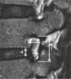
Percutaneous endoscopic lumbar foraminotomy (ELF) is a novel minimally invasive technique used to treat lumbar foraminal stenosis. However, the validity of foraminal decompression based on quantitative assessment using magnetic resonance imaging (MRI) has not yet been established. The objective of this study was to investigate the radiographic efficiency of ELF using MRI. Radiographic changes of neuroforamen were measured based on pre- and postoperative MRI findings. Images were blindly analyzed by two observers for foraminal stenosis grade and foraminal dimensions. The intraclass correlation coefficient (ICC) and k statistic were calculated to determine interobserver agreement. Thirty-five patients with 40 neuroforamen were evaluated. The mean visual analog scale (VAS) score improved from 8.4 to 2.1, and the mean Oswestry disability index (ODI) improved from 65.9 to 19.2. Overall, 91.4% of the patients achieved good or excellent outcomes. The mean grade of foraminal stenosis significantly improved from 2.63 to 0.68. There were significant increases in the mean foraminal area (FA) from 50.05 to 92.03 mm2, in mean foraminal height (FH) from 11.36 to 13.47 mm, in mean superior foraminal width (SFW) from 6.43 to 9.27 mm, and in mean middle foraminal width (MFW) from 1.47 to 78 mm (P < 0.001). Interobserver agreements for preoperative and postoperative measurements were good to excellent with the exception of SFW. In conclusion, foraminal dimensions and grades of foraminal stenosis significantly improved after ELF. These findings may enhance the clinical relevance of endoscopic lumbar foraminal decompression.
Keywords: endoscopic; foraminotomy; interobserver agreement; magnetic resonance imaging.
Conflict of interest statement
The authors declare no conflicts of interest.
Figures


References
-
- Reulen HJ, Pfaundler S, Ebeling U: The lateral microsurgical approach to the “extracanalicular” lumbar disc herniation. I: A technical note. Acta Neurochir (Wien) 84: 64–67, 1987 - PubMed
-
- Wiltse LL, Spencer CW: New uses and refinements of the paraspinal approach to the lumbar spine. Spine 13: 696–706, 1988 - PubMed
-
- Chang SB, Lee SH, Ahn Y, Kim JM: Risk factor for unsatisfactory outcome after lumbar foraminal and far lateral microdecompression. Spine 31: 1163–1167, 2006 - PubMed
-
- Kunogi J, Hasue M: Diagnosis and operative treatment of intraforaminal and extraforaminal nerve root compression. Spine 16: 1312–1320, 1991 - PubMed
-
- Donaldson WF, Star MJ, Thorne RP: Surgical treatment for the far lateral herniated lumbar disc. Spine 18: 1263–1267, 1993 - PubMed
MeSH terms
LinkOut - more resources
Full Text Sources
Other Literature Sources
Medical
Miscellaneous

