

The MARCKS protein amount is differently regulated by calpain during toxic effects of methylmercury between SH-SY5Y and EA.hy926 cells
- PMID: 29046508
- PMCID: PMC5745167
- DOI: 10.1292/jvms.17-0473
The MARCKS protein amount is differently regulated by calpain during toxic effects of methylmercury between SH-SY5Y and EA.hy926 cells
Abstract
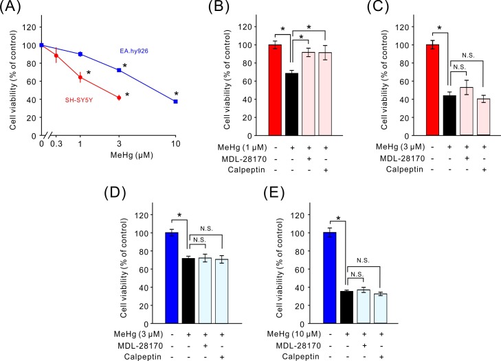
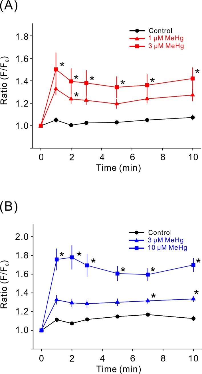
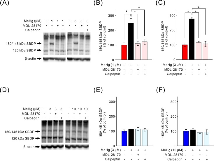
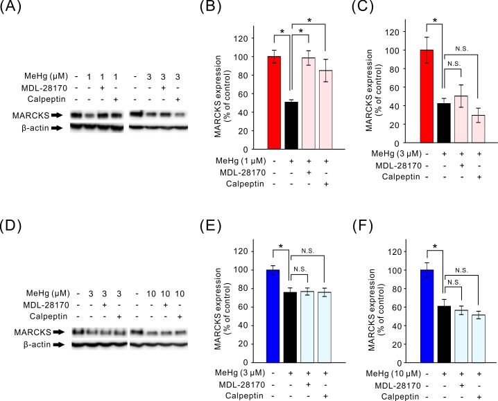
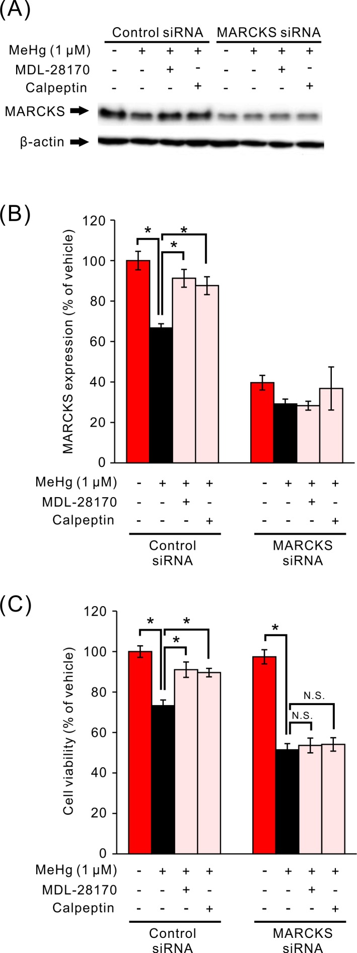
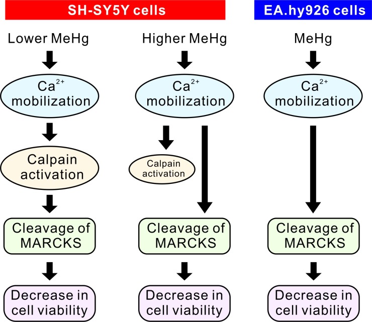
Methylmercury (MeHg) is an environmental pollutant that shows severe toxicity to humans and animals. However, the molecular mechanisms mediating MeHg toxicity are not completely understood. We have previously reported that the MARCKS protein is involved in the MeHg toxicity to SH-SY5Y neuroblastoma and EA.hy926 vascular endothelial cell lines. In addition, calpain, a Ca2+-dependent protease, is suggested to be associated with the MeHg toxicity. Because MARCKS is known as a substrate of calpain, we studied the relation between calpain activation and cleavage of MARCKS and its role in MeHg toxicity. In SH-SY5Y cells, MeHg decreased cell viability along with increased calcium mobilization, calpain activation and a decrease in MARCKS amounts. However, pretreatment with calpain inhibitors attenuated the decrease in cell viability and MARCKS amount induced only by 1 µM but not by 3 µM MeHg. In cells with a MARCKS knockdown, calpain inhibitors failed to attenuate the decrease in cell viability caused by MeHg. In EA.hy926 cells, although MeHg caused calcium mobilization and a decrease in MARCKS levels, calpain activation was not observed. These results indicate that the participation of calpain in the regulation of MARCKS amounts is dependent on the cell type and concentration of MeHg. In SH-SY5Y cells, calpain-mediated proteolysis of MARCKS is involved in cytotoxicity induced by a low concentration of MeHg.
Keywords: MARCKS; calpain; methylmercury.
Figures
Similar articles
-
MARCKS is involved in methylmercury-induced decrease in cell viability and nitric oxide production in EA.hy926 cells.J Vet Med Sci. 2016 Nov 1;78(10):1569-1576. doi: 10.1292/jvms.16-0249. Epub 2016 Jun 24. J Vet Med Sci. 2016. PMID: 27349763 Free PMC article.
-
Alteration in MARCKS phosphorylation and expression by methylmercury in SH-SY5Y cells and rat brain.Environ Toxicol Pharmacol. 2014 May;37(3):1256-63. doi: 10.1016/j.etap.2014.04.025. Epub 2014 Apr 28. Environ Toxicol Pharmacol. 2014. PMID: 24835554
-
ENaC activity is regulated by calpain-2 proteolysis of MARCKS proteins.Am J Physiol Cell Physiol. 2017 Jul 1;313(1):C42-C53. doi: 10.1152/ajpcell.00244.2016. Epub 2017 May 3. Am J Physiol Cell Physiol. 2017. PMID: 28468944 Free PMC article.
-
Low-dose methylmercury-induced oxidative stress, cytotoxicity, and tau-hyperphosphorylation in human neuroblastoma (SH-SY5Y) cells.Environ Toxicol. 2012 Sep;27(9):549-55. doi: 10.1002/tox.20672. Epub 2011 Jan 20. Environ Toxicol. 2012. PMID: 21254321
-
Calpain and MARCKS protein regulation of airway mucin secretion.Pulm Pharmacol Ther. 2012 Dec;25(6):427-31. doi: 10.1016/j.pupt.2012.06.003. Epub 2012 Jun 16. Pulm Pharmacol Ther. 2012. PMID: 22710197 Free PMC article.
Cited by
-
A peptide against the N-terminus of myristoylated alanine-rich C kinase substrate promotes neuronal differentiation in SH-SY5Y human neuroblastoma cells.J Vet Med Sci. 2024 Nov 1;86(11):1136-1144. doi: 10.1292/jvms.24-0276. Epub 2024 Sep 27. J Vet Med Sci. 2024. PMID: 39343539 Free PMC article.
-
Merits and Limitations of Studying Neuronal Depolarization-Dependent Processes Using Elevated External Potassium.ASN Neuro. 2020 Jan-Dec;12:1759091420974807. doi: 10.1177/1759091420974807. ASN Neuro. 2020. PMID: 33256465 Free PMC article. Review.
References
-
- Biamonte F., Latini L., Giorgi F. S., Zingariello M., Marino R., De Luca R., D’Ilio S., Majorani C., Petrucci F., Violante N., Senofonte O., Molinari M., Keller F.2014. Associations among exposure to methylmercury, reduced Reelin expression, and gender in the cerebellum of developing mice. Neurotoxicology 45: 67–80. doi: 10.1016/j.neuro.2014.09.006 - DOI - PubMed
-
- Castoldi A. F., Onishchenko N., Johansson C., Coccini T., Roda E., Vahter M., Ceccatelli S., Manzo L.2008. Neurodevelopmental toxicity of methylmercury: Laboratory animal data and their contribution to human risk assessment. Regul. Toxicol. Pharmacol. 51: 215–229. doi: 10.1016/j.yrtph.2008.03.005 - DOI - PubMed
MeSH terms
Substances
LinkOut - more resources
Full Text Sources
Other Literature Sources
Miscellaneous