. 2017 Oct 14:23:740-752.
eCollection 2017.

Elucidating the molecular basis of PPCD: Effects of decreased ZEB1 expression on corneal endothelial cell function

Affiliations

Elucidating the molecular basis of PPCD: Effects of decreased ZEB1 expression on corneal endothelial cell function

Marina Zakharevich et al. Mol Vis. .

Abstract

Purpose: To investigate the functional role that the zinc e-box binding homeobox 1 (ZEB1) gene, which underlies the genetic basis of posterior polymorphous corneal dystrophy 3 (PPCD3), plays in corneal endothelial cell proliferation, apoptosis, migration, and barrier function.

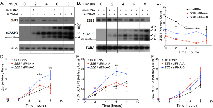
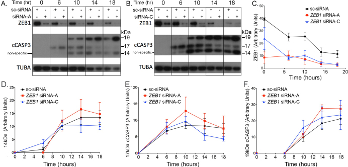
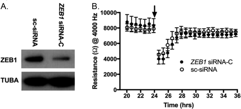
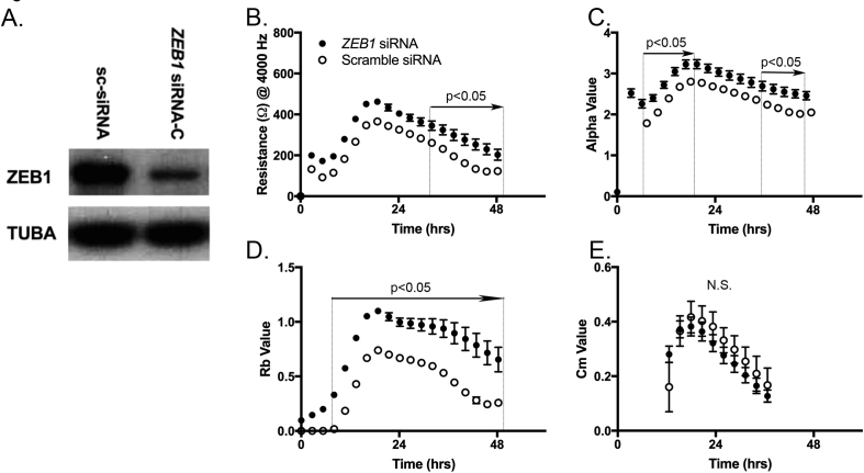
Methods: A human corneal endothelial cell line (HCEnC-21T) was transfected with siRNA targeting ZEB1 mRNA. Cell proliferation, apoptosis, migration, and barrier assays were performed: Cell proliferation was assessed with cell counting using a hemocytometer; cell apoptosis, induced by either ultraviolet C (UVC) radiation or doxorubicin treatment, was quantified by measuring cleaved caspase 3 (cCASP3) protein levels; and cell migration and barrier function were monitored with electric cell-substrate impedance sensing (ECIS).

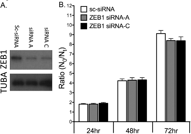
Results: ZEB1 knockdown in HCEnC-21T cells transfected with siRNA targeting ZEB1 did not result in a significant difference in cell proliferation when compared with control. Although knockdown of ZEB1 in HCEnC-21T cells sensitized the cells to UV-induced apoptosis, ZEB1 knockdown did not alter the cells' susceptibility to doxorubicin-induced apoptosis, as measured with cCASP3 protein levels, compared with controls. Similarly, no difference was observed in cell migration following ZEB1 knockdown. However, cell barrier function increased significantly following ZEB1 knockdown.

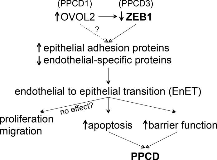
Conclusions: The corneal endothelium in PPCD3 is characterized by morphologic, anatomic, and molecular features that are more consistent with an epithelial-like rather than an endothelial-like phenotype. Although these characteristics have been well documented, we demonstrate for the first time that susceptibility to UV-induced apoptosis and cell barrier function are significantly altered in the setting of reduced ZEB1. The significance of an altered cellular response to apoptotic stimuli and increased cell barrier function in the pathobiology of PPCD remains to be fully elucidated.


Figures

Figure 1
Figure 1
Cell proliferation in HCEnC-21T cells transfected with siRNA. Cells were transfected with scrambled siRNA (sc-siRNA), ZEB1 siRNA-A, or ZEB1 siRNA-C. A: ZEB1 knockdown was demonstrated by western blotting for ZEB1, with TUBA used as a loading control. B: Cell quantities at 24, 48, and 72 h (Nt) were compared to the initial quantity (N0), and plotted as the ratio Nt/N0. Results represent 12 independent data points (n=12). Error bars = standard error of the mean (SEM).
Figure 2
Figure 2
Quantification of UV-induced apoptosis in ZEB1 knockdown cells. A, B: HCEnC-21T cells were transfected with ZEB1 siRNA-A, ZEB1 siRNA-C, or scrambled siRNA (sc-siRNA) and exposed to ultraviolet (UV) light for 0, 2, 4, 6, and 8 h. Apoptosis was assessed by the appearance of cleaved caspase 3 (cCASP3) with western blotting. C: ZEB1 knockdown was assessed with western blotting, quantified, and graphed. Abundance of the 14 kDa (D; non-specific), 17 kDa (E; active), and 19 kDa (F) fragments of cCASP3 was quantified with western blotting and graphed. Results represent three independent experiments (n = 3). Error bars = standard error of the mean (SEM), and **p<0.01 and ***p<0.001.
Figure 3
Figure 3
Quantification of DOX-induced apoptosis in ZEB1 knockdown cells. A, B: HCEnC-21T cells were transfected with ZEB1 siRNA-A, ZEB1 siRNA-C, or scrambled siRNA (sc-siRNA), and exposed to DOX for 0, 6, 10, 14, and 18 h. Apoptosis was assessed by the appearance of cleaved caspase 3 (cCASP3) with western blotting. C: ZEB1 knockdown was assessed with western blotting, quantified, and graphed. Abundance of the 14 kDa (D; non-specific), 17 kDa (E; active), and 19 kDa (F) fragments of cCASP3 was quantified with western blotting and graphed. Results represent three independent experiments (n = 3). Error bars = standard error of the mean (SEM).
Figure 4
Figure 4
The impact of ZEB1 reduction on cell migration in HCEnC-21T cells. A: HCEnC-21T cells were transfected with either ZEB1 siRNA-C or scrambled siRNA (sc-siRNA), and knockdown of ZEB1 was validated with western blotting. B: Transfected HCEnC-21T cells were seeded to 100% confluence on an electric cell–substrate impedance sensing (ECIS) electrode array. A high current (arrow) was applied to the circular electrode underneath the central region of the cell monolayer, thus clearing the circular area. Migration of cells into the cleared area was monitored for 12 h after wounding by measuring impedance (ohms) across the electrode at 4,000 Hz. Three wells for each condition were measured; n = 3. Error bars = standard error of the mean (SEM).
Figure 5
Figure 5
The impact of ZEB1 reduction on cell barrier function in in HCEnC-21T cells. A: HCEnC-21T cells were transfected with ZEB1 siRNA-C or scrambled siRNA (sc-siRNA), and knockdown of ZEB1 was confirmed with western blotting. B: Impedance (ohm) at 4,000 Hz, interendothelial resistance (Rb), cell attachment resistance (alpha), and plasma membrane capacitance (Cm) were measured for 48 h after seeding on electric cell–substrate impedance sensing (ECIS) electrode arrays. Three wells for each condition were measured; n = 3. Error bars = standard error of the mean (SEM). Statistical significance (p<0.05) is noted for a range when two or more sequential time points demonstrated statistical significance.
Figure 6
Figure 6
Hypothetical model of the mechanism underlying the progression of genotype to phenotype in posterior polymorphous corneal dystrophy.

References

    1. Krachmer JH. Posterior polymorphous corneal dystrophy: a disease characterized by epithelial-like endothelial cells which influence management and prognosis. Trans Am Ophthalmol Soc. 1985;83:413–75. - PMC - PubMed
    1. Weiss JS, Moller HU, Aldave AJ, Seitz B, Bredrup C, Kivela T, Munier FL, Rapuano CJ, Nischal KK, Kim EK, Sutphin J, Busin M, Labbe A, Kenyon KR, Kinoshita S, Lisch W. IC3D classification of corneal dystrophies–edition 2. Cornea. 2015;34:117–59. - PubMed
    1. Liskova P, Gwilliam R, Filipec M, Jirsova K, Reinstein Merjava S, Deloukas P, Webb TR, Bhattacharya SS, Ebenezer ND, Morris AG, Hardcastle AJ. High prevalence of posterior polymorphous corneal dystrophy in the Czech Republic; linkage disequilibrium mapping and dating an ancestral mutation. PLoS One. 2012;7:e45495. - PMC - PubMed
    1. Aldave AJ, Han J, Frausto RF. Genetics of the corneal endothelial dystrophies: an evidence-based review. Clin Genet. 2013;84:109–19. - PMC - PubMed
    1. Davidson AE, Liskova P, Evans CJ, Dudakova L, Noskova L, Pontikos N, Hartmannova H, Hodanova K, Stranecky V, Kozmik Z, Levis HJ, Idigo N, Sasai N, Maher GJ, Bellingham J, Veli N, Ebenezer ND, Cheetham ME, Daniels JT, Thaung CM, Jirsova K, Plagnol V, Filipec M, Kmoch S, Tuft SJ, Hardcastle AJ. Autosomal-Dominant Corneal Endothelial Dystrophies CHED1 and PPCD1 Are Allelic Disorders Caused by Non-coding Mutations in the Promoter of OVOL2. Am J Hum Genet. 2016;98:75–89. - PMC - PubMed

Publication types

MeSH terms

Supplementary concepts