

Impaired synaptic function is linked to cognition in Parkinson's disease
- PMID: 29046879
- PMCID: PMC5634342
- DOI: 10.1002/acn3.446
Impaired synaptic function is linked to cognition in Parkinson's disease
Abstract
Objective: Cognitive impairment is frequent in Parkinson's disease, but the underlying mechanisms are insufficiently understood. Because cortical metabolism is reduced in Parkinson's disease and closely associated with cognitive impairment, and CSF amyloid-β species are reduced and correlate with neuropsychological performance in Parkinson's disease, and amyloid-β release to interstitial fluid may be related to synaptic activity; we hypothesize that synapse dysfunction links cortical hypometabolism, reduced CSF amyloid-β, and presynaptic deposits of α-synuclein. We expect a correlation between hypometabolism, CSF amyloid-β, and the synapse related-markers CSF neurogranin and α-synuclein.
Methods: Thirty patients with mild-to-moderate Parkinson's disease and 26 healthy controls underwent a clinical assessment, lumbar puncture, MRI, 18F-fludeoxyglucose-PET, and a neuropsychological test battery (repeated for the patients after 2 years).
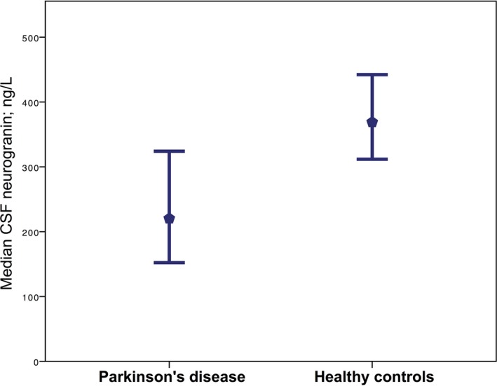
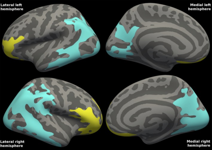
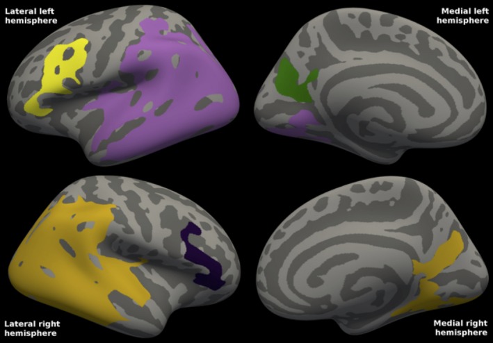
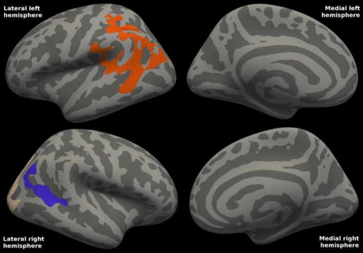
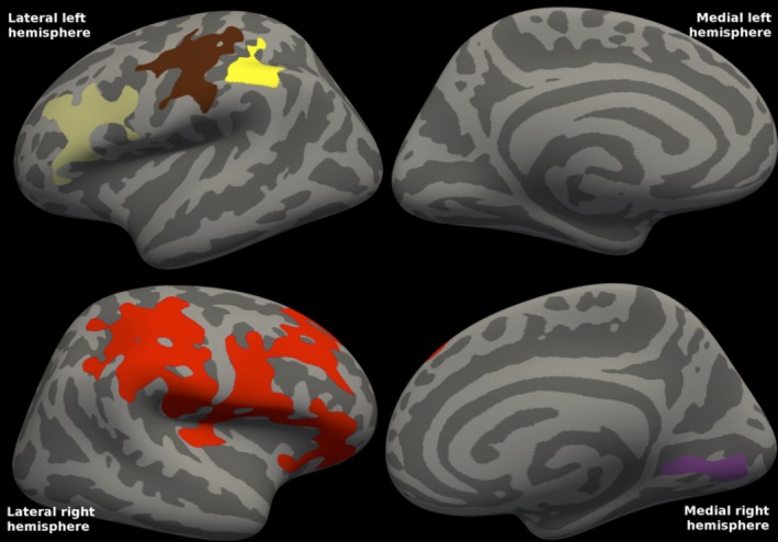
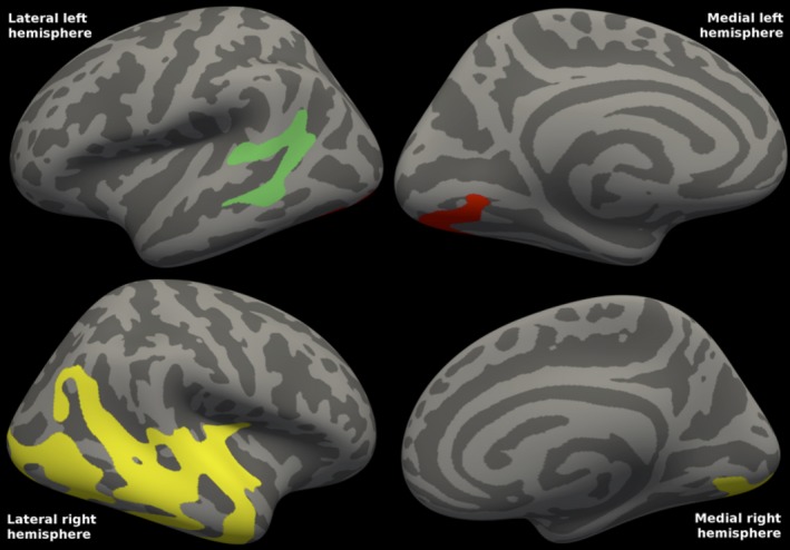
Results: All subjects had CSF amyloid-β 1-42 within normal range. In Parkinson's disease, we found strong significant correlations between cortical glucose metabolism, CSF Aβ, α-synuclein, and neurogranin. All PET CSF biomarker-based cortical clusters correlated strongly with cognitive parameters. CSF neurogranin levels were significantly lower in mild-to-moderate Parkinson's disease compared to controls, correlated with amyloid-β and α-synuclein, and with motor stage. There was little change in cognition after 2 years, but the cognitive tests that were significantly different, were also significantly associated with cortical metabolism. No such correlations were found in the control group.
Interpretation: CSF Aβ, α-synuclein, and neurogranin concentrations are related to cortical metabolism and cognitive decline. Synaptic dysfunction due to Aβ and α-synuclein dysmetabolism may be central in the evolution of cognitive impairment in Parkinson's disease.
Figures
Similar articles
-
Amyloid-β and α-synuclein cerebrospinal fluid biomarkers and cognition in early Parkinson's disease.Parkinsonism Relat Disord. 2015 Jul;21(7):758-64. doi: 10.1016/j.parkreldis.2015.04.027. Epub 2015 May 2. Parkinsonism Relat Disord. 2015. PMID: 25971633
-
Associations between Cerebrospinal Fluid Biomarkers and Cognition in Early Untreated Parkinson's Disease.J Parkinsons Dis. 2015;5(4):783-92. doi: 10.3233/JPD-150682. J Parkinsons Dis. 2015. PMID: 26599300 Free PMC article.
-
CSF β-Amyloid 1-42 Predicts Progression to Cognitive Impairment in Newly Diagnosed Parkinson Disease.J Mol Neurosci. 2016 Jan;58(1):88-92. doi: 10.1007/s12031-015-0647-x. Epub 2015 Sep 2. J Mol Neurosci. 2016. PMID: 26330275 Free PMC article.
-
Biological confounders for the values of cerebrospinal fluid proteins in Parkinson's disease and related disorders.J Neurochem. 2016 Oct;139 Suppl 1:290-317. doi: 10.1111/jnc.13390. Epub 2016 Feb 10. J Neurochem. 2016. PMID: 26452984 Review.
-
Cerebrospinal Fluid Amyloid β1-42, Tau, and Alpha-Synuclein Predict the Heterogeneous Progression of Cognitive Dysfunction in Parkinson's Disease.J Mov Disord. 2016 May;9(2):89-96. doi: 10.14802/jmd.16017. Epub 2016 May 25. J Mov Disord. 2016. PMID: 27240810 Free PMC article. Review.
Cited by
-
Proteomic Profiling of the Substantia Nigra to Identify Determinants of Lewy Body Pathology and Dopaminergic Neuronal Loss.J Proteome Res. 2021 May 7;20(5):2266-2282. doi: 10.1021/acs.jproteome.0c00747. Epub 2021 Apr 26. J Proteome Res. 2021. PMID: 33900085 Free PMC article.
-
Fluid markers of synapse degeneration in synucleinopathies.J Neural Transm (Vienna). 2022 Feb;129(2):187-206. doi: 10.1007/s00702-022-02467-8. Epub 2022 Feb 11. J Neural Transm (Vienna). 2022. PMID: 35147800 Review.
-
The disease-specific structural pattern in Parkinson's disease and its cortical characteristics associated with gene function: a 7-Tesla MRI study.J Neurol. 2025 Mar 31;272(4):300. doi: 10.1007/s00415-025-13035-x. J Neurol. 2025. PMID: 40159562
-
Pathophysiology of neurodegenerative diseases: An interplay among axonal transport failure, oxidative stress, and inflammation?Semin Immunol. 2022 Jan;59:101628. doi: 10.1016/j.smim.2022.101628. Epub 2022 Jun 30. Semin Immunol. 2022. PMID: 35779975 Free PMC article. Review.
-
CSF α-synuclein correlates with CSF neurogranin in late-life depression.Int J Neurosci. 2021 Apr;131(4):357-361. doi: 10.1080/00207454.2020.1744596. Epub 2020 Mar 31. Int J Neurosci. 2021. PMID: 32228205 Free PMC article.
References
-
- Aarsland D, Bronnick K, Fladby T. Mild cognitive impairment in Parkinson's disease. Curr Neurol Neurosci Rep 2011;11:371–378. - PubMed
-
- Aarsland D, Brønnick K, Larsen JP, et al. Cognitive impairment in incident, untreated Parkinson disease: the Norwegian ParkWest study. Neurology 2009;72:1121–1126. - PubMed
-
- Pienaar IS, Burn D, Morris C, Dexter D. Synaptic protein alterations in Parkinson's disease. Mol Neurobiol 2012;45:126–143. - PubMed
LinkOut - more resources
Full Text Sources
Other Literature Sources
