. 2017 Aug 30;4(10):722-738.
doi: 10.1002/acn3.450. eCollection 2017 Oct.

NMNAT3 is protective against the effects of neonatal cerebral hypoxia-ischemia

Affiliations

NMNAT3 is protective against the effects of neonatal cerebral hypoxia-ischemia

Rafael Galindo et al. Ann Clin Transl Neurol. .

Abstract

Objective: To determine whether the NAD+ biosynthetic protein, nicotinamide mononucleotide adenylyltransferase-3 (NMNAT3), is a neuroprotective inducible enzyme capable of decreasing cerebral injury after neonatal hypoxia-ischemia (H-I) and reducing glutamate receptor-mediated excitotoxic neurodegeneration of immature neurons.

Methods: Using NMNAT3-overexpressing mice we investigated whether increases in brain NMNAT3 reduced cerebral tissue loss following H-I. We then employed biochemical methods from injured neonatal brains to examine the inducibility of NMNAT3 and the mechanism of NMNAT3-dependent neuroprotection. Using AAV8-mediated vectors for in vitro neuronal NMNAT3 knockdown, we then examine the endogenous role of this protein on immature neuronal survival prior and following NMDA receptor-mediated excitotoxicity.

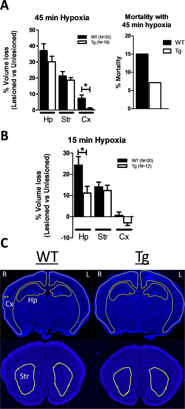
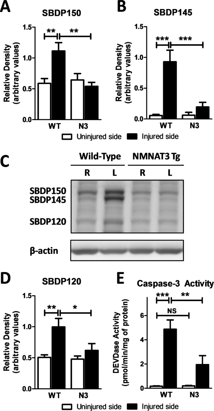
Results: NMNAT3 mRNA and protein levels increased after neonatal H-I. In addition, NMNAT3 overexpression decreased cortical and hippocampal tissue loss 7 days following injury. We further show that the NMNAT3 neuroprotective mechanism involves a decrease in calpastatin degradation, and a decrease in caspase-3 activity and calpain-mediated cleavage. Conversely, NMNAT3 knockdown of cortical and hippocampal neurons in vitro caused neuronal degeneration and increased excitotoxic cell death. The neurodegenerative effects of NMNAT3 knockdown were counteracted by exogenous upregulation of NMNAT3.

Conclusions: Our observations provide new insights into the neuroprotective mechanisms of NMNATs in the injured developing brain, adding NMNAT3 as an important neuroprotective enzyme in neonatal H-I via inhibition of apoptotic and necrotic neurodegeneration. Interestingly, we find that endogenous NMNAT3 is an inducible protein important for maintaining the survival of immature neurons. Future studies aimed at uncovering the mechanisms of NMNAT3 upregulation and neuroprotection may offer new therapies against the effects of hypoxic-ischemic encephalopathy.


Figures

Figure 1
Figure 1
Immunohistochemical localization of NMNAT3 in the young NMNAT3‐overexpressing postnatal brain. (A) NMNAT3 DAB immunohistochemistry of representative brain coronal sections from a NMNAT3 wild‐type (NMNAT3‐WT) and NMNAT3‐overexpressing transgenic (NMNAT3‐Tg) mouse brain at postnatal day 14. (B–D) Composite digital micrographs of NMNAT3 fluorescence immunoreactivity (NMNAT3‐IR) in the postnatal NMNAT3‐Tg hippocampus. NMNAT3‐IR is primarily found colocalized to neurons and regions of enriched neuronal processes as demonstrated by double immunofluorescence of NMNAT3 with neuronal antigen (B; NeuN) and neurofilament 200 (C and D; NF‐200). Scale bar = 110 μm (B), 440 μm (C), and 100 μm (D).
Figure 2
Figure 2
Endogenous NMNAT3 is induced by neonatal cerebral hypoxia and ischemia. (A) Bar graphs demonstrating the relative increase in NMNAT3 mRNA (from noninjured age‐matched controls) in the neonatal hypoxic (nonligated side) and hypoxic‐ischemic (H‐I) cerebral cortex (Cx) and hippocampus (Hp) 1, 3, and 7 days after injury at postnatal day 7 (number inside bar equals # of animals per condition). (B) NMNAT3 Western blot immunoreactivity (IR) of the neonatal hippocampus (Hp) from noninjured NMNAT3‐Tg (Tg) and wild‐type (WT) animals, and from WT hippocampi examined 24 h post‐H‐I. We consistently observed no NMNAT3 IR in control hippocampi. However, NMNAT3 immunoreactivity was notably present following injury in the hypoxic (Hypoxic Hp) and hypoxic‐ischemic hippocampus (H‐I Hp). Bar graph represents the relative quantitative density of the blots shown from the injured hippocampi relative to noninjured brains (N = 5). * p < 0.05; ** p < 0.01; *** p < 0.001
Figure 3
Figure 3
NMNAT3 overexpression reduces cortical and hippocampal tissue loss following neonatal H‐I. (A) Relative tissue loss (calculated by the percent difference between left (L) ipsilateral, ischemic, and right (R) contralateral, nonischemic, hemisphere) in the hippocampus (Hp), striatum (Str), and cerebral cortex (Cx) 7 days after carotid ligation followed by 45 min of hypoxia. Right bar graph shows total mortality during or immediately after 45 min of hypoxia chamber in wild‐type (WT) and NMNAT3 transgenic (Tg) mice. (B) Relative tissue loss 7 days post‐H‐I using 15 min of hypoxia chamber exposure, a paradigm with zero hypoxia‐induce mortality. (C) Representative coronal digital micrographs taken from injured WT and NMNAT3 Tg animals with 15 min of global hypoxia. * p < 0.05
Figure 4
Figure 4
NMNAT3 overexpression reduced the activation of calpain and caspase‐3 following neonatal H‐I in neonatal hippocampus. Quantitation (A, B, and D) and representative blot (C) of the relative immunoreactive band density by the corresponding spectrin breakdown product (SBPD; 145 and 150 kDa cleaved by calpain, 120 KDa cleaved by caspase) from wild‐type (WT) and NMNAT3‐overexpressing transgenic (N3) mice in the right (R) uninjured, nonischemic, versus left (L) injured, ischemic, hippocampus 24 h after H‐I. (E). Quantitation of caspase‐3 activity in WT and N3 transgenic mice of the injured and uninjured side measured 24 h after neonatal H‐I. N = 8 animals per group. * p < 0.05; ** p < 0.01; *** p < 0.001
Figure 5
Figure 5
NMNAT3 overexpression decreases the degradation of calpastatin following H‐I. Quantitation (A) and representative blot (B) of the relative calpastatin (CASTN) total band immunoreactive density by Western blot from wild‐type (WT) and NMNAT3‐overexpressing transgenic (N3) mice in the right (R) uninjured, nonischemic, versus left (L) injured, ischemic, hippocampus 24 h after H‐I (N = 8/group). ** p < 0.01; *** p < 0.001
Figure 6
Figure 6
AAV8 shRNA‐mediated GFP expression and relative gene expression of NMNATs in naïve, noninfected, and shNMNAT3 and/or human NMNAT3‐infected cortical neurons. (A) Quantitative amount of endogenous mouse NMNAT3 mRNA (Endog. N3; N = 4) versus viral‐transduced NMNAT3 mRNA in the presence (N = 3) or absence (N = 4) of AAV8‐mouse shNMNAT3 knockdown vector for 6 days. (B) Effect of AAV8‐mouse shNMNAT3 knockdown vector exposure on endogenous mRNA levels of mouse NMNAT1 (mN1), NMNAT2 (mN2), and NMNAT3 (mN3) in cortical neurons (N = 4 animals; each value calculated as average mRNA of three culture wells/animal/condition; negative value represents decrease from baseline). (C) Representative low‐power (bar = 440 μm) and high‐power (squared inserts; bar = 55 μm) single and composite micrographs of cultured cortical neurons stained for neuronal antigen (NeuN) 3 days after AAV8‐shNMNAT3‐GFP or AAV8‐shScramble‐GFP vector exposure. AAV8‐shNMNAT3 and shScramble vector expression is present in the majority of neurons and their processes by 3 days postinfection as assessed by GFP fluorescence. * p < 0.05; ** p < 0.01; *** p < 0.001
Figure 7
Figure 7
NMNAT3 mRNA depletion increases markers of neuronal cell death and enhances glutamate‐dependent excitotoxic degeneration of cortical neurons in vitro. The prodegenerative effect of NMNAT3 mRNA knockdown is reduced by NMNAT3 overexpression. (A) Effect of endogenous NMNAT3 knockdown on LDH activity in cortical neuronal cultures exposed to AAV8‐shNMNAT3‐GFP (shN3) virus for 6 days prior (N = 6) and 24 h (N = 4) after 50 μmol/L ibotenic acid exposure (shN3 + IBO), or exposed to a human NMNAT3‐overexpressing vector alone (N3; N = 6). The percent change in LDH activity was calculated as the change in LDH activity from that of experimentally paired neurons exposed to control AAV8‐shScramble vector for 6 days prior and 24 h after IBO exposure. Notice the significant elevation in LDH activity in shN3‐exposed neurons above shScramble control at baseline and following IBO exposure. LDH activity in N3‐exposed neuronal cultures appeared to be mildly reduced compared to shScramble controls. (B and C) Percent change in baseline LDH activity compared to shScramble controls (B) and amount of MAP2 immunoreactivity per unit area (C) 9 days postinfection with the mouse NMNAT3 knockdown AAV8 (shN3) in the presence (N = 4/group) or absence (N = 12/group) of the human NMNAT3‐overexpressing vector at the time (shN3 + N3), 3 days prior (N3 then shN3), or 3 days after (shN3 then N3) shN3 viral vector exposure. (D) Representative micrographs of shNMNAT3 GFP‐positive neurons alone (high‐power; bar = 110 μm) or stained with MAP2 (low‐power; bar = 1100 μm) and infected with the shN3 vector with or without N3‐overexpressing virus. GFP fragmentation, increased visualization of morphologically appearing astrocytes (see GFP‐shN3 panel), and a decrease in MAP2 + neurite staining was found primarily in shN3‐exposed neurons. The presence of the N3‐overexpressing vector in shN3‐treated neurons did not decrease, but rather qualitatively increase, shNMNAT3 GFP‐positive fluorescence.* p < 0.05; ** p < 0.01; *** p < 0.001
Figure 8
Figure 8
NMNAT3 mRNA knockdown also decreases hippocampal neuronal survival markers, an effect that is partially reversed by NMNAT3 upregulation. (A) Percent change in LDH activity compared to AAV8‐shScramble‐GFP viral control in hippocampal neurons infected with AAV8‐human NMNAT3 (N3; N = 6) or AAV8‐shNMNAT3‐GFP (shN3) virus for 6 days prior (N = 6) or 24 h (N = 4) after 50 μmol/L ibotenic acid exposure (IBO). Similar to the effect seen in cortical neurons, shN3 viral vector exposure increased LDH activity above control neuronal cultures prior and after injury with IBO. (B and C) Percent change in baseline LDH (B) and amount of MAP2 immunoreactivity per unit area (C) 9 days postneuronal infection with the mouse shN3 vector alone or 3 days after human N3 overexpressing vector (N3 then shN3; N = 6/group). (D) Representative micrographs of shNMNAT3‐GFP‐positive neurons (high‐power; bar = 110 μm) or stained with MAP2 (low‐power; bar = 1100 μm) and infected with the shN3 vector 3 days after N3‐overexpressing vector exposure. * p < 0.05; ** p < 0.01; *** p < 0.001

Similar articles

Cited by

References

    1. Ferriero DM. Neonatal brain injury. N Engl J Med 2004;351:1985–1995. - PubMed
    1. Higgins RD, Raju T, Edwards AD, et al. Hypothermia and other treatment options for neonatal encephalopathy: an executive summary of the Eunice Kennedy Shriver NICHD workshop. J Pediatr 2011;159:851‐858e1. - PMC - PubMed
    1. Shankaran S, Pappas A, McDonald SA, et al. Childhood outcomes after hypothermia for neonatal encephalopathy. N Engl J Med 2012;366:2085–2092. - PMC - PubMed
    1. Vexler ZS, Ferriero DM. Molecular and biochemical mechanisms of perinatal brain injury. Semin Neonatol 2001;6:99–108. - PubMed
    1. Berger F, Lau C, Dahlmann M, Ziegler M. Subcellular compartmentation and differential catalytic properties of the three human nicotinamide mononucleotide adenylyltransferase isoforms. J Biol Chem 2005;280:36334–36341. - PubMed

LinkOut - more resources