

Characterization of Pulmonary Nodules Based on Features of Margin Sharpness and Texture
- PMID: 29047033
- PMCID: PMC6113151
- DOI: 10.1007/s10278-017-0029-8
Characterization of Pulmonary Nodules Based on Features of Margin Sharpness and Texture
Abstract
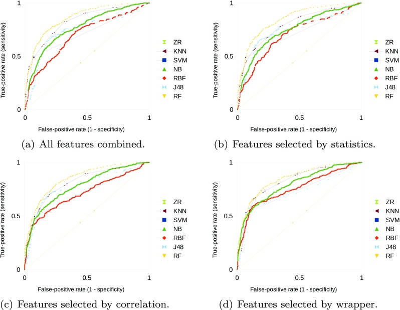
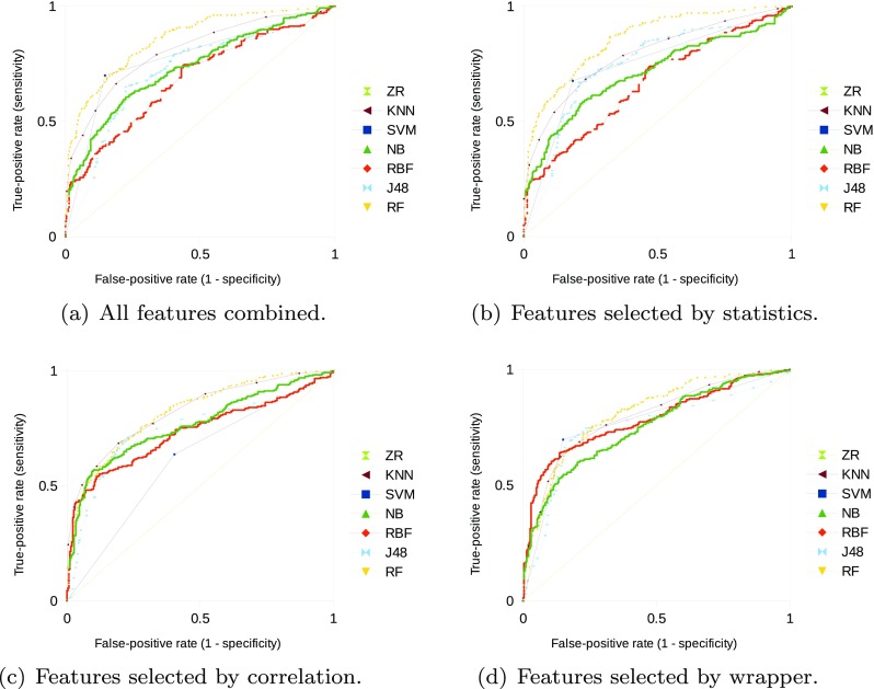
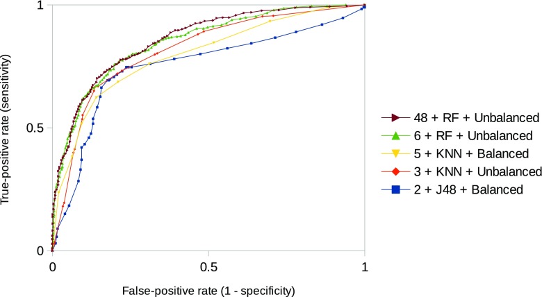
Lung cancer is the leading cause of cancer-related deaths in the world, and one of its manifestations occurs with the appearance of pulmonary nodules. The classification of pulmonary nodules may be a complex task to specialists due to temporal, subjective, and qualitative aspects. Therefore, it is important to integrate computational tools to the early pulmonary nodule classification process, since they have the potential to characterize objectively and quantitatively the lesions. In this context, the goal of this work is to perform the classification of pulmonary nodules based on image features of texture and margin sharpness. Computed tomography scans were obtained from a publicly available image database. Texture attributes were extracted from a co-occurrence matrix obtained from the nodule volume. Margin sharpness attributes were extracted from perpendicular lines drawn over the borders on all nodule slices. Feature selection was performed by different algorithms. Classification was performed by several machine learning classifiers and assessed by the area under the receiver operating characteristic curve, sensitivity, specificity, and accuracy. Highest classification performance was obtained by a random forest algorithm with all 48 extracted features. However, a decision tree using only two selected features obtained statistically equivalent performance on sensitivity and specificity.
Keywords: Image classification; Lung cancer; Pattern recognition; Pulmonary nodule.
Conflict of interest statement
Conflict of interests
The authors declare that they have no conflict of interest.
Ethical approval
For this type of study formal consent is not required. This study used a public image database (10.7937/K9/TCIA.2015.LO9QL9SX), which all protected health information (PHI) contained within the DICOM headers of the images were removed in accordance with Health Insurance Portability and Accountability Act (HIPAA) guidelines.
Figures
Similar articles
-
Selecting relevant 3D image features of margin sharpness and texture for lung nodule retrieval.Int J Comput Assist Radiol Surg. 2017 Mar;12(3):509-517. doi: 10.1007/s11548-016-1471-7. Epub 2016 Aug 23. Int J Comput Assist Radiol Surg. 2017. PMID: 27553081
-
Preoperative diagnosis of malignant pulmonary nodules in lung cancer screening with a radiomics nomogram.Cancer Commun (Lond). 2020 Jan;40(1):16-24. doi: 10.1002/cac2.12002. Epub 2020 Mar 3. Cancer Commun (Lond). 2020. PMID: 32125097 Free PMC article.
-
Artificial intelligence for detecting small FDG-positive lung nodules in digital PET/CT: impact of image reconstructions on diagnostic performance.Eur Radiol. 2020 Apr;30(4):2031-2040. doi: 10.1007/s00330-019-06498-w. Epub 2019 Dec 10. Eur Radiol. 2020. PMID: 31822970
-
Computed tomography and the secrets of lung nodules.Can Assoc Radiol J. 2015 Feb;66(1):2-4. doi: 10.1016/j.carj.2014.12.002. Can Assoc Radiol J. 2015. PMID: 25623006 Review. No abstract available.
-
Imaging of Solid Pulmonary Nodules.Clin Chest Med. 2024 Jun;45(2):249-261. doi: 10.1016/j.ccm.2023.08.013. Epub 2023 Sep 19. Clin Chest Med. 2024. PMID: 38816086 Review.
Cited by
-
Value of Shape and Texture Features from 18F-FDG PET/CT to Discriminate between Benign and Malignant Solitary Pulmonary Nodules: An Experimental Evaluation.Diagnostics (Basel). 2020 Sep 15;10(9):696. doi: 10.3390/diagnostics10090696. Diagnostics (Basel). 2020. PMID: 32942729 Free PMC article.
-
An Appraisal of Nodule Diagnosis for Lung Cancer in CT Images.J Med Syst. 2019 May 15;43(7):181. doi: 10.1007/s10916-019-1327-0. J Med Syst. 2019. PMID: 31093830
-
The Effects of Perinodular Features on Solid Lung Nodule Classification.J Digit Imaging. 2021 Aug;34(4):798-810. doi: 10.1007/s10278-021-00453-2. Epub 2021 Mar 31. J Digit Imaging. 2021. PMID: 33791910 Free PMC article.
-
CT-based radiomics for prediction of histologic subtype and metastatic disease in primary malignant lung neoplasms.Int J Comput Assist Radiol Surg. 2020 Jan;15(1):163-172. doi: 10.1007/s11548-019-02093-y. Epub 2019 Nov 13. Int J Comput Assist Radiol Surg. 2020. PMID: 31722085
-
Form Factors as Potential Imaging Biomarkers to Differentiate Benign vs. Malignant Lung Lesions on CT Scans.Sensors (Basel). 2022 Jul 4;22(13):5044. doi: 10.3390/s22135044. Sensors (Basel). 2022. PMID: 35808538 Free PMC article.
References
-
- Wu H, Sun T, Wang J, Li X, Wang W, Huo D, Lv P, He W, Wang K, Guo X. Combination of radiological and gray level co-occurrence matrix textural features used to distinguish solitary pulmonary nodules by computed tomography. J Digit Imaging. 2013;26:797–802. doi: 10.1007/s10278-012-9547-6. - DOI - PMC - PubMed
-
- Aerts HJ, Velazquez ER, Leijenaar RT, Parmar C, Grossmann P, Cavalho S, Bussink J, Monshouwer R, Haibe-Kains B, Rietveld D, Hoebers F, Rietbergen MM, Leemans CR, Dekker A, Quackenbush J, Gillies RJ, Lambin P. Decoding tumour phenotype by noninvasive imaging using a quantitative radiomics approach. Nat Commun. 2014;5:4006. doi: 10.1038/ncomms5006. - DOI - PMC - PubMed
-
- Awai K, Murao K, Ozawa A, Nakayama Y, Nakaura T, Liu D, Kawanaka K, Funama Y, Morishita S, Yamashita Y. Pulmonary nodules: estimation of malignancy at thin-section helical CT—effect of computer-aided diagnosis on performance of radiologists. Radiology. 2006;239:276–284. doi: 10.1148/radiol.2383050167. - DOI - PubMed
Publication types
MeSH terms
LinkOut - more resources
Full Text Sources
Other Literature Sources
Medical
