. 2017:967:105-137.
doi: 10.1007/978-3-319-63245-2_8.

ROS Signaling in the Pathogenesis of Acute Lung Injury (ALI) and Acute Respiratory Distress Syndrome (ARDS)

Affiliations

ROS Signaling in the Pathogenesis of Acute Lung Injury (ALI) and Acute Respiratory Distress Syndrome (ARDS)

Manuela Kellner et al. Adv Exp Med Biol. 2017.

Abstract

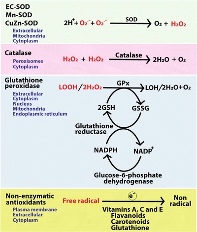
The generation of reactive oxygen species (ROS) plays an important role for the maintenance of cellular processes and functions in the body. However, the excessive generation of oxygen radicals under pathological conditions such as acute lung injury (ALI) and its most severe form acute respiratory distress syndrome (ARDS) leads to increased endothelial permeability. Within this hallmark of ALI and ARDS, vascular microvessels lose their junctional integrity and show increased myosin contractions that promote the migration of polymorphonuclear leukocytes (PMNs) and the transition of solutes and fluids in the alveolar lumen. These processes all have a redox component, and this chapter focuses on the role played by ROS during the development of ALI/ARDS. We discuss the origins of ROS within the cell, cellular defense mechanisms against oxidative damage, the role of ROS in the development of endothelial permeability, and potential therapies targeted at oxidative stress.

Keywords: Catalase; Cytochrome P450; Glutathione; Lung injury; Mitochondrial respiratory chain; NADPH oxidase; Nitric oxide synthase; Polymorphonuclear leukocytes; Pulmonary endothelial cell; Reactive oxygen species; Superoxide dismutase; Xanthine oxidase.


Figures

Fig. 1
Fig. 1
Dysfunction of microvascular endothelium and alveolar epithelium in ARDS. Polymorphonuclear leukocytes (PMNs) and macrophages infiltrate the inflamed region through the microvascular blood vessels releasing cytotoxic factors such as pro-inflammatory cytokines and ROS. Theese cytokines and ROS contribute to the endothelial and epithelial dysfunction resulting in leakage of fluids from circulation into the interstitial space and alveoli. This results in pulmonary edema and impaired gas exchange. Sources of inflammation range from bacterial infections to mechanical ventilation
Fig. 2
Fig. 2
Sources of reactive oxygen species. Mitochondria, NADPH oxidase, xanthine oxidase, and eNOS are the major contributors of ROS in cells of vasculature during active metabolism. NADPH oxidase in phagocytic cells such as macrophages and neutrophils that are resident in blood vessels contribute to a significant amount of superoxide (O2 ·−). Endothelial NOS (eNOS) generates NO free radicals that interact with O2 ·− to generate peroxynitrite. Peroxynitrite induces nitrasative stress on cells by nitrating proteins and altering signaling pathways. When eNOS is uncoupled, it can generate superoxide. Oxidative phosphorylations in mitochondria are a source of O2 ·−. Especially complexes I, III, and IV generate O2 ·− when there is a leak of electrons at subsequent transfer stages. O2 ·− generated in mitochondria is often immediately dismutated to H2O2 by SOD which can cross mitochondrial membrane as well as cell membranes. Other lesser sources of ROS are cytochrome P450 enzymes which often generate O2 ·− during detoxification of xenobiotics and they are predominantly expressed in hepatic tissue. ADP adenosine diphosphate, ATP adenosine triphosphate, BH 4 tetrahydrobiopterin, Ca-Calmodulin calcium and calmodulin, CoQ coenzyme Q, Cyt c cytochrome c, eNOS endothelial nitric oxide synthase, FADH 2 flavin adenine dinucleotide, H 2 O 2 hydrogen peroxide, IMM inner mitochondrial membrane, IMS inter-mitochondrial membrane space, NADH nicotinamide adenine dinucleotide, NADPH nicotinamide adenine dinucleotide phosphate, NO nitric oxide, OMM outer mitochondrial membrane, Pi inorganic phosphate, O 2 superoxide free radical, ONOO peroxynitrite free radical, SOD superoxide dismutase, complex I—NADH oxidoreductase (I), complex II—succinate dehydrogenase (II), complex III—cytochrome c reductase (III), complex IV—cytochrome c oxidase (IV), complex V—ATP synthase (V), XH xenobiotic, XOH alcohol/aldehyde form of xenobiotic
Fig. 3.
Fig. 3.
The antioxidant system in cells. Enzymatic and nonenzymatic antioxidants catalyze reactions to neutralize free radicals by donating electrons. Enzymatic antioxidants catalyze reactions to neutralize specific free radicals such that superoxide dismutase (SOD) dismutates superoxide to hydrogen peroxide (H2O2), and catalase and glutathione peroxidase (GPx) convert hydrogen peroxide to water. GPx also converts lipid hyroperoxides (LOOH) to lipid alcohols or aldehydes (LOH). Glutathione reductase replenishes reduced glutathione (GSH) pools from oxidized glutathione (GSSG) using NADPH as reducing equivalents. Nonenzymatic antioxidants such as vitamins, flavonoids and glutathione can also reduce free radicals by donating electrons
Fig. 4.
Fig. 4.
Comparison of the transcellular and paracellular transport in physiological and pathological conditions. The transport of fluids, solutes, and macromolecules occur over transcellular and paracellular pathways. Under physiological conditions both transcellular and the paracellular transport are highly restricted, whereas under pathological conditions increased vascular permeability can be observed. ROS have several distinct impacts on the endothelial barrier. Initially, within the transcellular pathway caveolin-1 is affected by ROS leading to increased vascular permeability. ROS mainly influence the paracellular pathway through decreased expression and oligomerization of the junctional proteins as well as increases in the phosphorylation of junctional proteins on both serine and tyrosine residues. Both ROCE and SOCE are affected by ROS leading to increased endothelial Ca2+ influx. This increases calcium/calmodulin-dependent phosphorylation of myosin light chains leading to myosin contraction. Both ROCE and also SOCE are affected by ROS. AJs adherens junctions, cADPR cyclic adenosine triphosphate ribose, DAG diacylglycerol, ER endoplasmatic reticulum, GPCR G protein coupled receptor, IP 3 inositol triphosphate, JAMs junctional adhesion molecules, MLC myosin light chain, MLCK myosin light chain kinase, MLCP myosin light chain phosphatase, PLC phospholipase C, PKC protein kinase C, ROCE receptor-operated calcium entry, ROS reactive oxygen species, SOCE store-operated calcium entry, TJ tight junctions, TRPC/M transient receptor potential canonical/melastatin, VOOs vesiculo-vacuolar organelles, ZO zonula occludens
Fig. 5.
Fig. 5.
ROS-mediated endothelial polymorphonuclear leukocyte migration. Over both transcellular and paracellular pathways, polymorphonuclear leukocytes (PMNs) pass through the endothelium to migrate from the blood lumen into the alveolar lumen. In both pathways selectin, integrins, and immunoglobulins (Ig) help facilitate this cellular migration. The transcellular migration of PMNs through the cell body is a rare event. More commonly paracellular migration occurs. This requires a number of migration steps: (1) tethering and rolling; (2) activation; (3) adhesion; (4) crawling; (5) transendothelial migration, and (6) diapedesis. Within each migration steps, varying cell adhesion molecules (CAMs) are needed. Selectins modulate the initially tethering and rolling of PMNs on the inner surface of the blood vessel, whereby PMNs start to slow down. Based on the rolling, endothelial cells are activated to release chemokines (CXCL1 or CXCL8) that transmit and bind to chemokine receptors (CXCLR1 and CXCLR8) localized on the surface of the PMNs. This causes PMN localized integrins to change from a low-affinity state (inactive bent conformation) to a high-affinity state (active fully extended conformation) forming a firm adhesion to CAMs of the Ig superfamily. The ultimate entry of the PMNs into the blood barrier occurs over these established CAMs formations. Increased ROS leads to enhanced invasion of immune cells into the injured tissue. ROS also regulate the expression CAMs both directly and through transcription factors (NF-κB, AP-1) that exert major influences on PMN migration. NF-κB nuclear factor-kappa-B, PMNs polymorphonuclear leukocytes, ROS reactive oxygen species

Similar articles

Cited by

References

    1. Ashbaugh DG, Bigelow DB, Petty TL, Levine BE. Acute respiratory distress in adults. Lancet (London, England) 1967;2(7511):319–323. doi: 10.1016/S0140-6736(67)90168-7. - DOI - PubMed
    1. Ranieri VM, Rubenfeld GD, Thompson BT, Ferguson ND, Caldwell E, Fan E, et al. Acute respiratory distress syndrome: The berlin definition. Journal of the American Medical Association. 2012;307(23):2526–2533. - PubMed
    1. Matthay MA, Wiener-Kronish JP. Intact epithelial barrier function is critical for the resolution of alveolar edema in humans. The American Review of Respiratory Disease. 1990;142(6 Pt 1):1250–1257. doi: 10.1164/ajrccm/142.6_Pt_1.1250. - DOI - PubMed
    1. Gropper MA, Wiener-Kronish J. The epithelium in acute lung injury/acute respiratory distress syndrome. Current Opinion in Critical Care. 2008;14(1):11–15. doi: 10.1097/MCC.0b013e3282f417a0. - DOI - PubMed
    1. Ware LB, Matthay MA. Alveolar fluid clearance is impaired in the majority of patients with acute lung injury and the acute respiratory distress syndrome. American Journal of Respiratory and Critical Care Medicine. 2001;163(6):1376–1383. doi: 10.1164/ajrccm.163.6.2004035. - DOI - PubMed

MeSH terms